Review
. 2011 Jan 20;13(1):9.
doi: 10.1186/1532-429X-13-9.

Repaired tetralogy of Fallot: the roles of cardiovascular magnetic resonance in evaluating pathophysiology and for pulmonary valve replacement decision support

Affiliations
Review

Repaired tetralogy of Fallot: the roles of cardiovascular magnetic resonance in evaluating pathophysiology and for pulmonary valve replacement decision support

Tal Geva. J Cardiovasc Magn Reson. .

Abstract

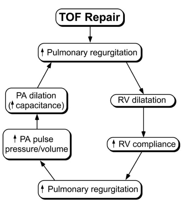
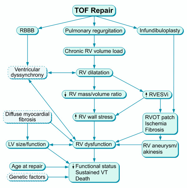
Surgical management of tetralogy of Fallot (TOF) results in anatomic and functional abnormalities in the majority of patients. Although right ventricular volume load due to severe pulmonary regurgitation can be tolerated for many years, there is now evidence that the compensatory mechanisms of the right ventricular myocardium ultimately fail and that if the volume load is not eliminated or reduced by pulmonary valve replacement the dysfunction might be irreversible. Cardiovascular magnetic resonance (CMR) has evolved during the last 2 decades as the reference standard imaging modality to assess the anatomic and functional sequelae in patients with repaired TOF. This article reviews the pathophysiology of chronic right ventricular volume load after TOF repair and the risks and benefits of pulmonary valve replacement. The CMR techniques used to comprehensively evaluate the patient with repaired TOF are reviewed and the role of CMR in supporting clinical decisions regarding pulmonary valve replacement is discussed.


Figures

Figure 1
Figure 1
Some of the factors influencing progression of pulmonary regurgitation after TOF repair. This process likely plateaus at a certain point but the time course of PR has not been fully characterized.
Figure 2
Figure 2
Steady-state free precession cine CMR of the right ventricular (RV) inflow and outflow showing a large outflow tract patch aneurysm (An). RA = right atrium
Figure 3
Figure 3
Correlation between pulmonary regurgitation and right ventricular (RV) end-diastolic volume index in 206 patients with repaired TOF [46].
Figure 4
Figure 4
Relationship between right ventricular end-systolic volume and RV ejection fraction in 100 patients with repaired TOF (Spearman rank correlation coefficient (rs) = -0.77; p < 0.001) [10].
Figure 5
Figure 5
Three-dimensional surface models of the right ventricular (RV) free wall reconstructed from multi-slice 2-dimensional short- and long-axis images. Top panel: Scar tissue map based on late gadolinium enhancement (LGE) imaging showing extensive late hyperenhancement of the RVOT (yellow and orange). Bottom panel: Displacement map based on multi-slice cine SSFP showing dyskinesis of the RVOT (red). See Wald et al. for further technical details [42].
Figure 6
Figure 6
Factors influencing right ventricular (RV) dysfunction and impaired clinical status after TOF repair.
Figure 7
Figure 7
Association between right ventricular (RV) and left ventricular (LV) ejection fraction (EF) in 100 patients with repaired TOF [10].
Figure 8
Figure 8
CMR imaging in a 37 year-old patient with repaired TOF, severe pulmonary regurgitation, moderate tricuspid regurgitation (arrow), and severe right ventricular dilatation (end-diastolic volume index 386 ml/m2) and dysfunction (ejection fraction 15%). This patient who also exhibited severe heart failure symptoms had only modest decrease in RV size and no improvement in RV function after pulmonary valve replacement.
Figure 9
Figure 9
Evaluation of ventricular size and function by ECG-gated cine SSFP MR in repaired TOF: Left ventricular 2-chamber (vertical long-axis) plane.
Figure 10
Figure 10
Evaluation of ventricular size and function by ECG-gated cine SSFP MR in repaired TOF: Right ventricular 2-chamber (vertical long-axis) plane. RA = right atrium; RV = right ventricle; RVOT = right ventricular outflow tract.
Figure 11
Figure 11
Evaluation of biventricular size and function by ECG-gated cine SSFP MR in repaired TOF: 4-chamber (horizontal long-axis) plane.
Figure 12
Figure 12
Evaluation of biventricular size and function by ECG-gated cine SSFP MR in repaired TOF: Ventricular short-axis. Note that 16 short-axis slices were required to fully cover the markedly dilated RV in this patient.
Figure 13
Figure 13
Evaluation of the right ventricular outflow tract (RVOT) long-axis by ECG-gated cine SSFP MR. Along with the RV 2-chamber plane (Figure 10), this view demonstrates patency of the RVOT and main pulmonary artery, presence or absence of pulmonary valve tissue, and wall motion abnormalities.
Figure 14
Figure 14
Gadolinium-enhanced 3-dimensional magnetic resonance angiography in a patient with repair TOF and a giant aneurysm of the outflow patch.
Figure 15
Figure 15
Evaluation of pulmonary regurgitation (PR) by ECG-gated cine phase contrast MR: The imaging plane is placed perpendicular to the long-axis of the main pulmonary artery (MPA).
Figure 16
Figure 16
Evaluation of pulmonary regurgitation (PR) by ECG-gated cine phase contrast MR: Color-coded flow map of the main pulmonary artery with the region of interest contour shown at peak systole.
Figure 17
Figure 17
Evaluation of pulmonary regurgitation (PR) by ECG-gated cine phase contrast MR: MPA flow rate (Y-axis) versus time (X-axis). Flow above the baseline represents antegrade flow and flow below the baseline represents retrograde (regurgitation) flow.
Figure 18
Figure 18
Late gadolinium enhancement imaging in the ventricular short-axis showing intense late hyperenhancement in the RVOT (white arrow) and weak hyperenhancement in the superior and inferior junctions between the interventricular septum and the free wall (yellow arrows). The former represents scar tissue and is associated with regional wall motion abnormalities. The latter is a commonly observed finding in patients with repaired TOF and its clinical importance is uncertain [42,85].
Figure 19
Figure 19
CMR assessment of biventricular volumes and mass in repaired TOF. Cross-referencing between ventricular long- and short-axis imaging planes aids determining inclusion of basal slices in the ventricular volume analysis. When an operator selects a frame on the short-axis grid, that location is highlighted on the linked horizontal and vertical long axis images, allowing the operator to determine the location of the slab relative to the atrioventricular valves. Right lower panel: Examples of contour drawings on the left and right ventricular endocardial and epicardial boundaries at the base, mid-ventricular, and apical levels.
Figure 20
Figure 20
Plot of left and right ventricular volumes versus time throughout the cardiac cycle in a patient with repaired TOF. Note that minimal RV volume occurs 70 ms (2 cardiac phases) after minimal LV volume.
Figure 21
Figure 21
Computer model of right ventricular stress map based on CMR data of a patient with repaired TOF. This and similar models include fluid-structure interactions, two-layer RV-LV structure, anisotropic material properties, fiber orientation, and active contraction [139,140].
Figure 22
Figure 22
Tissue tracking of the ventricular myocardium (LV = left panel; RV = right panel) in a patient with repaired TOF. Analysis of circumferential strain is performed in the ventricular short-axis plane using a commercial tissue tracking software package and custom-built filters to modify the DICOM headers of the CMR datasets to allow them to be analyzed by the software package. The myocardium is divided into 6 segments and circumferential strain (Y-axis;%) versus time (X-axis; milliseconds) is plotted for each segment. The time difference to peak circumferential strain--a measure of ventricular synchrony--measured 83 ms in the LV and 389 ms in the RV, reflecting RV dyssynchrony.

References

    1. Blalock A, Taussig HB. The surgical treatment of malformation of the heart in which there is pulmonary stenosis or pulmonary atresia. JAMA. 1945;128:189–202. - PubMed
    1. Lillehei CW, Cohen M, Warden HE, Varco RL. The direct-vision intracardiac correction of congenital anomalies by controlled cross circulation; results in thirty-two patients with ventricular septal defects, tetralogy of Fallot, and atrioventricularis communis defects. Surgery. 1955;38:11–29. - PubMed
    1. Lillehei CW, Cohen M, Warden HE, Read RC, Aust JB, Dewall RA, Varco RL. Direct vision intracardiac surgical correction of the tetralogy of Fallot, pentalogy of Fallot, and pulmonary atresia defects; report of first ten cases. Ann Surg. 1955;142:418–42. doi: 10.1097/00000658-195509000-00010. - DOI - PMC - PubMed
    1. Kirklin JW, Wallace RB, McGoon DC, DuShane JW. Early and late results after intracardiac repair of Tetralogy of Fallot. 5-Year review of 337 patients. Ann Surg. 1965;162:578–89. doi: 10.1097/00000658-196510000-00004. - DOI - PMC - PubMed
    1. Bacha EA, Scheule AM, Zurakowski D, Erickson LC, Hung J, Lang P, Mayer JE Jr, del Nido PJ, Jonas RA. Long-term results after early primary repair of tetralogy of Fallot. J Thorac Cardiovasc Surg. 2001;122:154–61. doi: 10.1067/mtc.2001.115156. - DOI - PubMed

Publication types

MeSH terms