. 2011 Apr;300(4):G538-46.
doi: 10.1152/ajpgi.00342.2010. Epub 2011 Jan 20.

The extracellular calcium-sensing receptor is required for cholecystokinin secretion in response to L-phenylalanine in acutely isolated intestinal I cells

Affiliations

The extracellular calcium-sensing receptor is required for cholecystokinin secretion in response to L-phenylalanine in acutely isolated intestinal I cells

Alice P Liou et al. Am J Physiol Gastrointest Liver Physiol. 2011 Apr.

Abstract

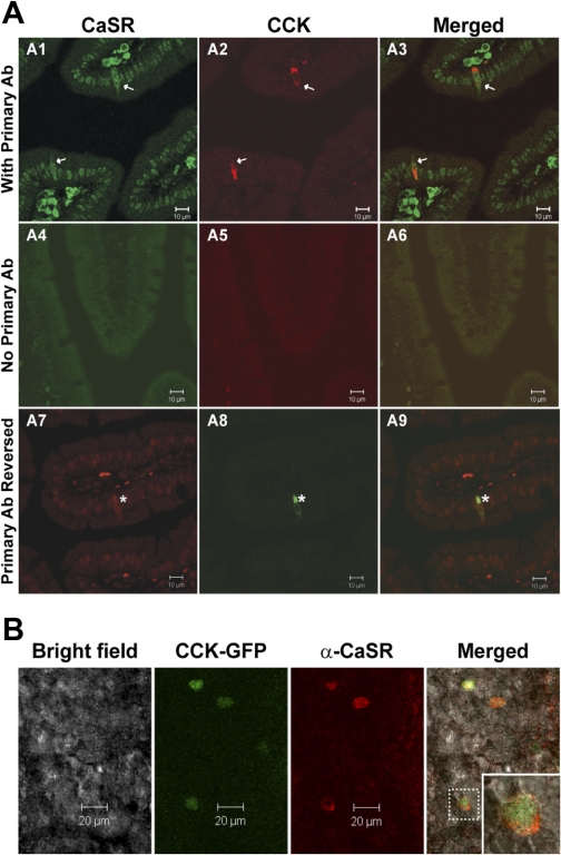
The extracellular calcium-sensing receptor (CaSR) has recently been recognized as an L-amino acid sensor and has been implicated in mediating cholecystokinin (CCK) secretion in response to aromatic amino acids. We investigated whether direct detection of L-phenylalanine (L-Phe) by CaSR results in CCK secretion in the native I cell. Fluorescence-activated cell sorting of duodenal I cells from CCK-enhanced green fluorescent protein (eGFP) transgenic mice demonstrated CaSR gene expression. Immunostaining of fixed and fresh duodenal tissue sections confirmed CaSR protein expression. Intracellular calcium fluxes were CaSR dependent, stereoselective for L-Phe over D-Phe, and responsive to type II calcimimetic cinacalcet in CCK-eGFP cells. Additionally, CCK secretion by an isolated I cell population was increased by 30 and 62% in response to L-Phe in the presence of physiological (1.26 mM) and superphysiological (2.5 mM) extracellular calcium concentrations, respectively. While the deletion of CaSR from CCK-eGFP cells did not affect basal CCK secretion, the effect of L-Phe or cinacalcet on intracellular calcium flux was lost. In fact, both secretagogues, as well as superphysiological Ca(2+), evoked an unexpected 20-30% decrease in CCK secretion compared with basal secretion in CaSR(-/-) CCK-eGFP cells. CCK secretion in response to KCl or tryptone was unaffected by the absence of CaSR. The present data suggest that CaSR is required for hormone secretion in the specific response to L-Phe by the native I cell, and that a receptor-mediated mechanism may inhibit hormone secretion in the absence of a fully functional CaSR.


Figures

Fig. 1.
Fig. 1.
Acute isolation of cholecystokinin (CCK)-enhanced green fluorescent protein (eGFP) (“I”) cells with fluorescent-activated cell sorting (FACS). Single-cell suspensions from the duodenal mucosa were analyzed and selected based on green fluorescent protein (GFP) fluorescence. Distinct non-eGFP (light gray) and eGFP (black) cell populations were visualized by flow cytometric analysis pre- (A) and postsorting (B). The P4 gating indicates the cutoff point between non-eGFP and eGFP cells. Postsorted eGFP-negative cells were determined to be dead cells by Trypan blue exclusion. C: all sorted eGFP-positive cells (green) were confirmed to be both GFP positive (blue) and CCK positive (red) by immunostaining. PE-A, phycoerythrin area; GFP-A, GFP area.
Fig. 2.
Fig. 2.
Gene expression of putative peptide or amino acid sensing receptor proteins in CCK-eGFP cells relative to non-eGFP cells. Gene transcripts for calcium-sensing receptor (CaSR) are significantly elevated in CCK-eGFP cells, whereas gene expression for PepT1, GPR92, and T1R3 are equivalent to that expressed in the non-eGFP cell population. T1R1 transcript was undetectable in both populations. Gene fold expression of 1 is regarded as equivalent to the non-eGFP population. Significance was determined using a paired t-test of the ΔCT (comparative threshold) values from 3–5 separate experimental preparations. N.D., not detectable. Values are means ± SE. ***P < 0.0001.
Fig. 3.
Fig. 3.
Immunohistochemical colocalization of CaSR on CCK-expressing enteroendocrine cells. A: CCK-immunoreactive enteroendocrine cells express CaSR (arrows, asterisks) in paraffin-embedded duodenal tissue sections of CaSR+/+ PTH+/+ mice. CCK immunolabeling was concentrated on the basilar aspect of isolated cells. CaSR-specific immunostaining was distinguished from the high background nonspecific staining of nuclei within these sections by visualizing whole cell CaSR immunostaining throughout all microscopic focal planes within CCK-immunopositive cells. Primary antibody (Ab) was excluded in tissue sections represented in A4A6. A1A3, with primary Ab; A7A9, primary Ab reversed. B: expression of CaSR in CCK-eGFP+ cells in fresh primary duodenal villi from CaSR+/+ CCK-eGFP transgenic mice. Inset: an enlarged CaSR-positive CCK-eGFP cell outlined in the villus demonstrating CaSR expression localized to the cell surface.
Fig. 4.
Fig. 4.
l- or d-Phenylalanine (Phe)-stimulated changes in intracellular Ca2+ in CaSR+/+ and CaSR−/− CCK-eGFP cells. A: effect of l- or d-Phe (20 mM) on Ca2+ responses in CCK-eGFP cells. Representative time courses for changes in Quest Rhod4 fluorescence intensity after stimulation with either l-Phe (♦) or d-Phe (◊) in CaSR+/+ (top) and CaSR−/− (bottom) CCK-eGFP cells are shown. Arrow indicates time of stimulation. B: l- or d-Phe (20 mM) induced increases in intracellular Ca2+ concentration ([Ca2+]i) in CaSR+/+ (solid bars) vs. CaSR−/− (open bars) CCK-eGFP cells. Changes in [Ca2+]i in CCK-eGFP cells were analyzed as maximum %increase in the fluorescence intensity (FI) of Quest Rhod4 relative to the baseline. Values are means ± SE of maximum %increase. Maximum %increase was calculated as follows: (peak FI − basal FI)/basal FI × 100. *P < 0.05, d-Phe for CaSR+/+ (n = 9) vs. CaSR−/− (n = 4); ***P < 0.001, l-Phe (n = 11) vs. d-Phe (n = 9), CaSR+/+; ****P < 0.0001, l-Phe, CaSR+/+ (n = 11) vs. CaSR−/− (n = 6), or l-Phe (n = 11) vs. 0 mM vehicle control (n = 4).
Fig. 5.
Fig. 5.
Effect of CaSR expression on stimulated secretion of CCK from isolated I cells. CCK secretion was from sorted CaSR+/+ (solid bars; A) and CaSR−/− (open bars; B) CCK-eGFP cells in response to a 30-min incubation with l-Phe (30 mM) alone and with additional extracellular Ca2+ (2.5 mM), 2.5 mM Ca2+ alone, d-Phe (30 mM), 1% tryptone, and KCl (50 mM). CCK secreted in the supernatant was measured by RIA. Results are presented as %change (means ± SE) from baseline CCK secretion in Hank's balanced salt solution supplemented with 10 mM HEPES (HHBSS) alone. N = 6–9 separate cell preparations for all treatments, with each condition performed and sampled in triplicate. *P < 0.05 and **P < 0.01, relative to baseline secretion within each genotype.
Fig. 6.
Fig. 6.
Effect of cinacalcet on intracellular Ca2+ flux and CCK secretion from I cells in the presence or absence of CaSR. A: representative time courses of changes in [Ca2+]i in Quest Rhod4-loaded CCK-eGFP cells stimulated with cinacalcet (1 μM). ♦, CaSR+/+; ◊, CaSR−/−; arrow, time of stimulation. B: mean maximum %increase (±SE) in the FI of Quest Rhod4 relative to the baseline [(peak FI − basal FI)/basal FI × 100] in CasR+/+ (n = 16) vs. CasR−/− (n = 10) CCK-eGFP cells after stimulation with cinacalcet, 1 μM. ****P < 0.0001 with unpaired t-test. C: CCK released in the supernatant of sorted CaSR+/+ (solid bar) and CaSR−/− (open bar) CCK-eGFP cells incubated with cinacalcet (1 μM). Results are presented as %change (means ± SE) from baseline secretion in 0.05% DMSO-HHBSS vehicle alone. N = 5–7 separate cell populations with each condition performed and sampled in triplicate. *P < 0.05.

Comment in

Similar articles

Cited by

References

    1. Anika SM, Houpt TR, Houpt KA. Satiety elicited by cholecystokinin in intact and vagotomized rats. Physiol Behav 19: 761– 766, 1977 - PubMed
    1. Ballinger AB, Clark ML. l-Phenylalanine releases cholecystokinin (CCK) and is associated with reduced food intake in humans: evidence for a physiological role of CCK in control of eating. Metabolism 43: 735– 738, 1994 - PubMed
    1. Bezencon C, le Coutre J, Damak S. Taste-signaling proteins are coexpressed in solitary intestinal epithelial cells. Chem Senses 32: 41– 49, 2007 - PubMed
    1. Bouras EP, Misukonis MA, Liddle RA. Role of calcium in monitor peptide-stimulated cholecystokinin release from perifused intestinal cells. Am J Physiol Gastrointest Liver Physiol 262: G791– G796, 1992 - PubMed
    1. Brown EM, Gamba G, Riccardi D, Lombardi M, Butters R, Kifor O, Sun A, Hediger MA, Lytton J, Hebert SC. Cloning and characterization of an extracellular Ca(2+)-sensing receptor from bovine parathyroid. Nature 366: 575– 580, 1993 - PubMed

Publication types

LinkOut - more resources