Review
. 2011 May;300(5):G684-96.
doi: 10.1152/ajpgi.00474.2010. Epub 2011 Jan 20.

Intestinal myofibroblasts: targets for stem cell therapy

Affiliations
Review

Intestinal myofibroblasts: targets for stem cell therapy

R C Mifflin et al. Am J Physiol Gastrointest Liver Physiol. 2011 May.

Abstract

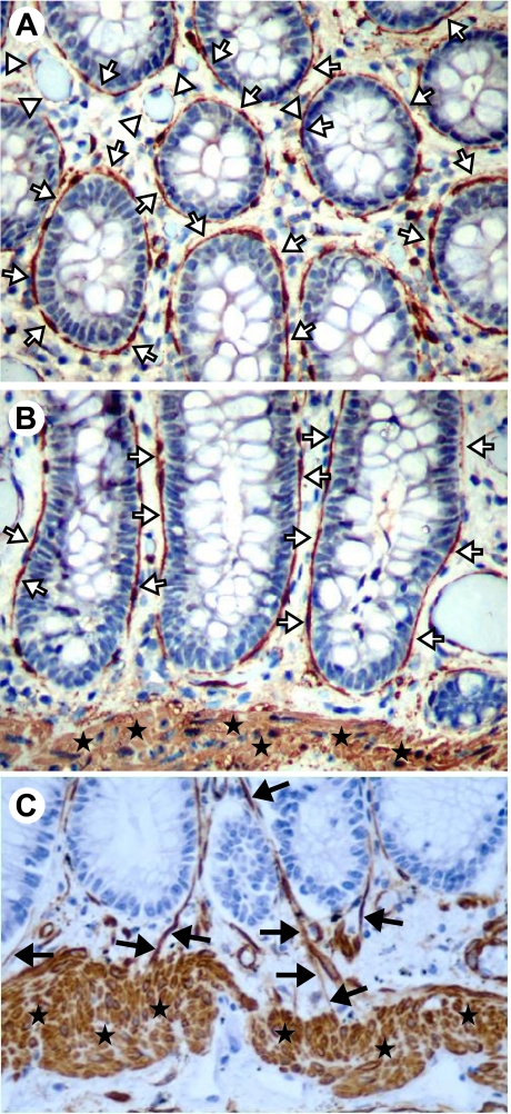
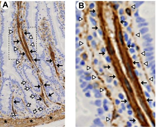
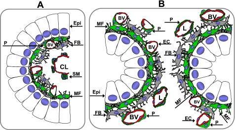
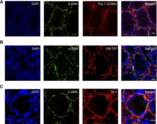
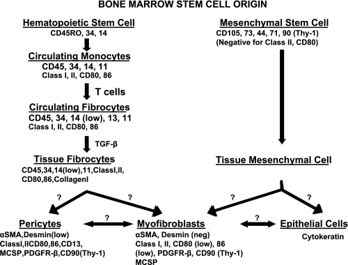
The subepithelial intestinal myofibroblast is an important cell orchestrating many diverse functions in the intestine and is involved in growth and repair, tumorigenesis, inflammation, and fibrosis. The myofibroblast is but one of several α-smooth muscle actin-positive (α-SMA(+)) mesenchymal cells present within the intestinal lamina propria, including vascular pericytes, bone marrow-derived stem cells (mesenchymal stem cells or hematopoietic stem cells), muscularis mucosae, and the lymphatic pericytes (colon) and organized smooth muscle (small intestine) associated with the lymphatic lacteals. These other mesenchymal cells perform many of the functions previously attributed to subepithelial myofibroblasts. This review discusses the definition of a myofibroblast and reconsiders whether the α-SMA(+) subepithelial cells in the intestine are myofibroblasts or other types of mesenchymal cells, i.e., pericytes. Current information about specific, or not so specific, molecular markers of lamina propria mesenchymal cells is reviewed, as well as the origins of intestinal myofibroblasts and pericytes in the intestinal lamina propria and their replenishment after injury. Current concepts and research on stem cell therapy for intestinal inflammation are summarized. Information about the stem cell origin of intestinal stromal cells may inform future stem cell therapies to treat human inflammatory bowel disease (IBD).


Figures

Fig. 1.
Fig. 1.
α-Smooth muscle actin (α-SMA)-positive cells of the colonic mucosa. Cross section (A) and longitudinal sections (B and C) of the colonic mucosa show positive (brown) α-SMA immunostaining of pericryptal (subepithelial) myofibroblasts (white arrows) and the muscularis mucosae (stars). Vascular and/or lymphatic pericytes (white arrowheads) are also visible. Original magnification ×200. C: pericytes surrounding lymphatics (black arrows) are seen entering the muscularis mucosae in this section. Note that the lamina propria blood vessels between the crypts and muscularis mucosae are also highlighted (original magnification ×400). [A and B taken from Powell et al. (96), with permission. C adapted from Adegboyega et al. (3), with permission].
Fig. 2.
Fig. 2.
α-SMA-positive cells of the small intestinal mucosa. A: longitudinal section of normal human jejunum with positive (brown) α-SMA immunostaining of myofibroblasts (white arrows), pericytes (white arrowheads), lymphatic lacteal-associated smooth muscle (black arrows), and muscularis mucosae (stars). B: magnified image of box shown in A demonstrating intensely α-SMA-positive smooth muscle (black arrows) associated with the lymphatic lacteal and mildly α-SMA-positive cells (white arrowheads) subjacent to the epithelium. These subepithelial cells appear to be predominantly pericytes rather than myofibroblasts. [Courtesy of Patrick Adegboyega, Louisiana State University Health Sciences Center (Shreveport, LA). Adapted from Powell et al. (99) with permission.]
Fig. 3.
Fig. 3.
Schematic depicting the spatial relationships of the epithelium and mesenchymal elements of the small intestinal villus and colonic crypts. A: cross section through a small intestinal villus showing the epithelium (Epi) and lamina propria. α-SMA+ subepithelial cells are predominantly pericytes (P, green) surrounding small blood vessels (BV). There may be occasional myofibroblasts (MF), although these could all be pericytes in the villus. There are smooth muscle (SM) bundles (dark green) associated with the central lymphatic lacteal (CL). Fibroblasts (FB, gray) are shown. B: cross section through the colonic crypts demonstrating relationships among the various mesenchymal elements in the lamina propria: subepithelial myofibroblasts (MF, green) and pericytes (P, green) surrounding blood vessels (BV) and lymphatics (L). Fibroblasts (F, gray) are found deeper in the lamina propria. EC, endothelial cell.
Fig. 4.
Fig. 4.
Identification of the intestinal mesenchymal cells. A–C: confocal microscopic analysis of multicolor immunofluorescent staining of cross sections of a normal human colonic crypt. CD90, ER-TR7, or TE-7 (red) identify nonhematopoietic mesenchymal cells of the colonic mucosa: fibroblasts, myofibroblasts, and pericytes of the small vessels and lymphatics. The subepithelial myofibroblast network and pericytes are identified on the basis of their morphology, subepithelial or perivascular location, and immunoreactivity to α-SMA (green) and mesenchymal markers CD90, ER-TR7, or TE-7 (red), resulting in orange-yellow color on the merged images. A: cell nuclei (blue) stained with DAPI; α-SMA shown in green; Thy-1 (CD90) shown in red. B: cell nuclei (blue) stained with DAPI; α-SMA shown in green; ER-TR7 shown in red. C: cell nuclei (blue) stained with DAPI; α-SMA shown in green; TE-7 shown in red. Calibration bars are 20 μm.
Fig. 5.
Fig. 5.
Bone marrow stem cell origin of intestinal mesenchymal stromal cells. Diagram demonstrates possible stem cell origin and routes for derivation of lamina propria mesenchymal cells (and epithelial cells) from bone marrow precursors. See text for discussion. TGF-β, transforming growth factor-β; MCSP, melanoma chondroitin sulfate proteoglycan; PDGFR-β, PDGF receptor-β.

References

    1. Abe R, Donnelly SC, Peng T, Bucala R, Metz CN. Peripheral blood fibrocytes: differentiation pathway and migration to wound sites. J Immunol 166: 7556–7562, 2001 - PubMed
    1. Adegboyega PA, Mifflin RC, DiMari JF, Saada JI, Powell DW. Immunohistochemical study of myofibroblasts in normal colonic mucosa, hyperplastic polyps, and adenomatous colorectal polyps. Arch Pathol Lab Med 126: 829–836, 2002 - PubMed
    1. Adegboyega PA, Ololade O, Saada J, Mifflin R, Di Mari JF, Powell DW. Subepithelial myofibroblasts express cyclooxygenase-2 in colorectal tubular adenomas. Clin Cancer Res 10: 5870–5879, 2004 - PubMed
    1. Aertgeerts K, Levin I, Shi L, Snell GP, Jennings A, Prasad GS, Zhang Y, Kraus ML, Salakian S, Sridhar V, Wijnands R, Tennant MG. Structural and kinetic analysis of the substrate specificity of human fibroblast activation protein alpha. J Biol Chem 280: 19441–19444, 2005 - PubMed
    1. Andoh A, Bamba S, Brittan M, Fujiyama Y, Wright NA. Role of intestinal subepithelial myofibroblasts in inflammation and regenerative response in the gut. Pharmacol Therapeut 114: 94–106, 2007 - PubMed

Publication types

LinkOut - more resources