. 2011 Mar 4;108(5):555-65.
doi: 10.1161/CIRCRESAHA.110.221911. Epub 2011 Jan 20.

Reactive oxygen species-activated Ca/calmodulin kinase IIδ is required for late I(Na) augmentation leading to cellular Na and Ca overload

Affiliations

Reactive oxygen species-activated Ca/calmodulin kinase IIδ is required for late I(Na) augmentation leading to cellular Na and Ca overload

Stefan Wagner et al. Circ Res. .

Abstract

Rationale: In heart failure Ca/calmodulin kinase (CaMK)II expression and reactive oxygen species (ROS) are increased. Both ROS and CaMKII can increase late I(Na) leading to intracellular Na accumulation and arrhythmias. It has been shown that ROS can activate CaMKII via oxidation.

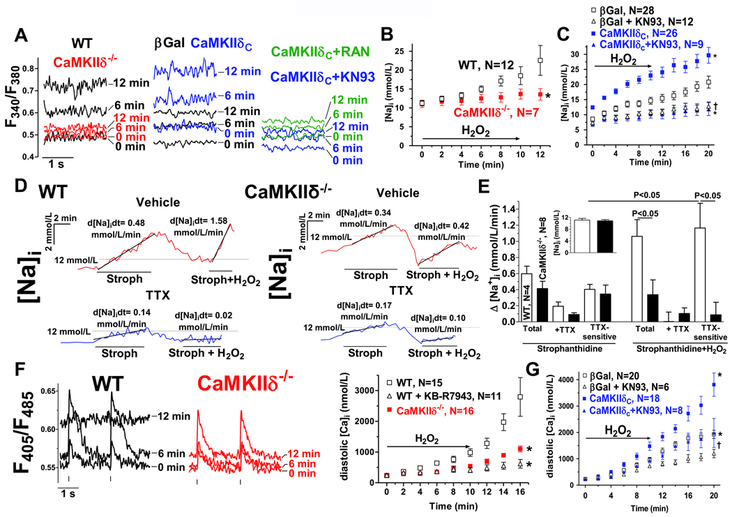
Objective: We tested whether CaMKIIδ is required for ROS-dependent late I(Na) regulation and whether ROS-induced Ca released from the sarcoplasmic reticulum (SR) is involved.

Methods and results: 40 μmol/L H(2)O(2) significantly increased CaMKII oxidation and autophosphorylation in permeabilized rabbit cardiomyocytes. Without free [Ca](i) (5 mmol/L BAPTA/1 mmol/L Br(2)-BAPTA) or after SR depletion (caffeine 10 mmol/L, thapsigargin 5 μmol/L), the H(2)O(2)-dependent CaMKII oxidation and autophosphorylation was abolished. H(2)O(2) significantly increased SR Ca spark frequency (confocal microscopy) but reduced SR Ca load. In wild-type (WT) mouse myocytes, H(2)O(2) increased late I(Na) (whole cell patch-clamp). This increase was abolished in CaMKIIδ(-/-) myocytes. H(2)O(2)-induced [Na](i) and [Ca](i) accumulation (SBFI [sodium-binding benzofuran isophthalate] and Indo-1 epifluorescence) was significantly slowed in CaMKIIδ(-/-) myocytes (versus WT). CaMKIIδ(-/-) myocytes developed significantly less H(2)O(2)-induced arrhythmias and were more resistant to hypercontracture. Opposite results (increased late I(Na), [Na](i) and [Ca](i) accumulation) were obtained by overexpression of CaMKIIδ in rabbit myocytes (adenoviral gene transfer) reversible with CaMKII inhibition (10 μmol/L KN93 or 0.1 μmol/L AIP [autocamtide 2-related inhibitory peptide]).

Conclusions: Free [Ca](i) and a functional SR are required for ROS activation of CaMKII. ROS-activated CaMKIIδ enhances late I(Na), which may lead to cellular Na and Ca overload. This may be of relevance in hear failure, where enhanced ROS production meets increased CaMKII expression.


Figures

Figure 1
Figure 1
ROS can activate CaMKII in permeabilized myocytes (free [Ca]i 20 nmol/L) Western blots of p-CaMKII (A) or ox-CaMKII (B) vs. CaMKII and GAPDH are shown. Mean densitometric values for p-CaMKII vs. CaMKII (C) or ox-CaMKII vs. CaMKII (D) are shown. One way ANOVA was used for statistical comparison. All densitometric values are summarized in Online Table I. CaMKII was stimulated with 1 µmol/L [Ca], 200 or 40 µmol/L H2O2. For some experiments, caffeine (10 mmol/L) and THA (5 µmol/L) were used to unload the SR or AIP (1 µmol/L) was present.
Figure 2
Figure 2
ROS activation of CaMKII is Ca-dependent. Permeabilized myocytes bathed in heavy Ca buffers (5 mmol/L BAPTA/1 mmol/L Br2-BAPTA) were used for Western blots of p-CaMKII (A) or ox-CaMKII (B). All densitometric values are shown in Online Table I.
Figure 3
Figure 3
Ca sparks were measured in permeabilized rabbit myocytes. A) Line scan images and typical corresponding Ca sparks. Mean data for CaSpF, Ca spark amplitude and FDHM (B) or calculated SR Ca leak (C) are shown. D) SR Ca content was estimated using caffeine (10 mmol/L).
Figure 4
Figure 4
Ca sparks were measured in intact twitching mouse myocytes (BCL 2 s). A) Line scan images at baseline and 4 min after onset of H2O2 (200 µmol/L) without any pharmacological intervention (left) and in the presence of KN93 (10 µmol/L, right). Mean data for CaSpF, amplitude and FDHM (B) and calculated SR Ca leak (C) are shown. D) SR Ca load (caffeine 10 mmol/L) is shown.
Figure 5
Figure 5
Late INa after exposure to H2O2. Late INa was measured at BCL 2 s (protocol in inset) in myocytes from CaMKIIδ−/− mice (vs. WT littermates) and in isolated rabbit myocytes after CaMKIIδC overexpression (vs. βGal). Late INa was measured as the integral between 50 and 500 ms after onset of depolarisation. Original traces (A) and mean data (B) showed that H2O2 significantly increased late INa in WT myocytes. This increase was completely abolished in myocytes lacking CaMKIIδ. Similarly, H2O2 significantly increased late INa in rabbit myocytes (βGal, C&D). CaMKIIδC overexpression markedly enhanced H2O2-induced late INa, and AIP (100 nmol/L) significantly blunted this increase. *P<0.05 (two-way ANOVA) vs. WT or βGal, respectively; †P<0.05 (two-way ANOVA) vs. CaMKIIδC.
Figure 6
Figure 6
[Na]i and [Ca]i upon exposure to H2O2. A) Original traces of SBFI fluorescence in mouse (left panel) and rabbit ventricular myocytes (mid and right panel). Data is shown for baseline (0 min), 6 and 12 min after onset of H2O2 exposition (200 µmol/L). WT–black, CaMKIIδ−/−–red, CaMKIIδC overexpression–blue, βGal–black, KN93–blue, RAN–green. Mean data after conversion to [Na]i is shown for mouse (B) and rabbit myocytes (C). D) Original traces of Na influx (via [Na]i-converted SBFI fluorescence) in mouse ventricular myocytes (BCL 2s) exposed to 100 µmol/L strophanthidin (Stroph) (with and without 1 µmol/L TTX and/or 200 µmol/L H2O2). E) Mean data for the rate of rise of [Na]i (baseline [Na]i in inset). (one-way ANOVA). F) Original traces of Indo-1 fluorescence at baseline (0 min), 6 and 12 min after onset of exposure to H2O2 (myocytes from WT, left panel and CaMKIIδ−/−, mid panel). Vertical bars indicate the electrical stimulus. Right panel shows mean data after conversion to [Ca]i. G) Mean data for rabbit myocytes. CaMKIIδC overexpression significantly enhanced (vs. βGal) and CaMKII inhibition (10 µmol/L KN93) significantly slowed the increase in diastolic [Ca]i after H2O2. *P<0.05 (two-way ANOVA) vs. WT or βGal, respectively, †P<0.05 (two-way ANOVA) vs. CaMKIIδC
Figure 7
Figure 7
ROS-induced arrhythmias. A) Original traces of stimulated APs before and after H2O2 treatment. Mean data shows a significant H2O2-dependent increase in AP duration (APD 90, B), and a significant positive shift in the diastolic action potential in WT (C), but no change in APD and no positive shift diastolic action potential in CaMKIIδC−/− myocytes. Insets show baseline APD 90 and diastolic potential, respectively. D) Original traces of consecutive APs (left) in myocytes from WT or CaMKIIδC−/− mice. Mean data (right) shows that H2O2-induces EADs+DADs were significantly more frequent in WT vs. CaMKIIδC−/−. E) Original traces of sarcomere length in intact twitching myocytes from WT or CaMKIIδC−/− mice. F) Mean data. The propensity for H2O2-induced arrhythmias was significantly reduced in CaMKIIδC−/−. G) Kaplan-Meier analysis of hypercontracture development. *P<0.05 vs. WT. BCL 2 s, vertical bars indicate stimuli.

References

    1. Tsutsui H, Ide T, Hayashidani S, Suematsu N, Utsumi H, Nakamura R, Egashira K, Takeshita A. Greater susceptibility of failing cardiac myocytes to oxygen free radical-mediated injury. Cardiovasc Res. 2001;49:103–109. - PubMed
    1. Kinugawa S, Tsutsui H, Hayashidani S, Ide T, Suematsu N, Satoh S, Utsumi H, Takeshita A. Treatment with dimethylthiourea prevents left ventricular remodeling and failure after experimental myocardial infarction in mice: Role of oxidative stress. Circ Res. 2000;87:392–398. - PubMed
    1. Mallat Z, Philip I, Lebret M, Chatel D, Maclouf J, Tedgui A. Elevated levels of 8-iso-prostaglandin f-2 alpha in pericardial fluid of patients with heart failure - a potential role for in vivo oxidant stress in ventricular dilatation and progression to heart failure. Circulation. 1998;97:1536–1539. - PubMed
    1. Josephson R, Silverman H, Lakatta E, Stern M, Zweier J. Study of the mechanisms of hydrogen-peroxide and hydroxyl free radical-induced cellular injury and calcium overload in cardiac myocytes. J Biol Chem. 1991;266:2354–2361. - PubMed
    1. Wagner S, Seidler T, Picht E, Maier LS, Kazanski V, Teucher N, Schillinger W, Pieske B, Isenberg G, Hasenfuss G, Kögler H. Na+- Ca2+ exchanger overexpression predisposes to reactive oxygen species-induced injury. Cardiovasc Res. 2003;60:404–412. - PubMed

Publication types

MeSH terms