. 2011 Mar 4;108(5):574-81.
doi: 10.1161/CIRCRESAHA.110.222844. Epub 2011 Jan 20.

Endothelial cell-specific deficiency of Ang II type 1a receptors attenuates Ang II-induced ascending aortic aneurysms in LDL receptor-/- mice

Affiliations

Endothelial cell-specific deficiency of Ang II type 1a receptors attenuates Ang II-induced ascending aortic aneurysms in LDL receptor-/- mice

Debra L Rateri et al. Circ Res. .

Abstract

Rationale: Human studies and mouse models have provided evidence for angiotensin II (Ang II)-based mechanisms as an underlying cause of aneurysms localized to the ascending aorta. In agreement with this associative evidence, we have published recently that Ang II infusion induces aneurysmal pathology in the ascending aorta.

Objective: The aim of this study was to define the role of angiotensin II type 1a (AT(1a)) receptors and their cellular location in Ang II-induced ascending aortic aneurysms (AAs).

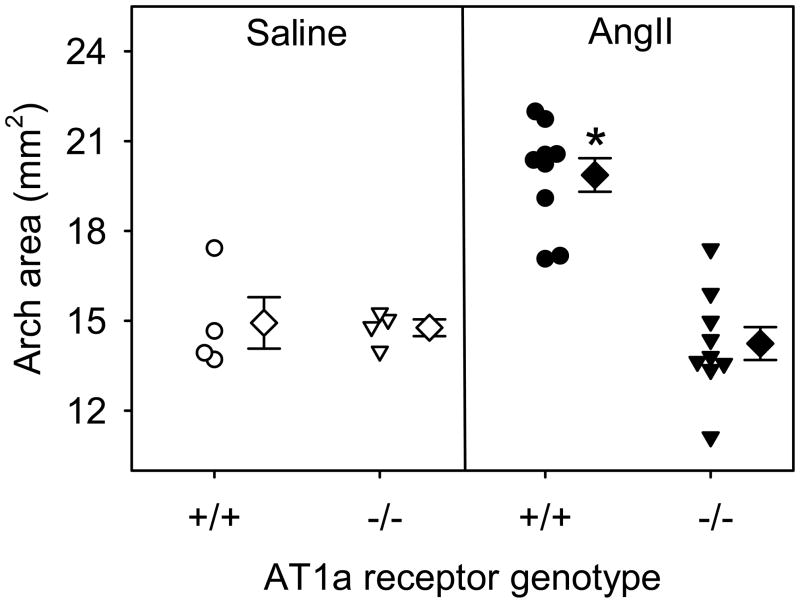
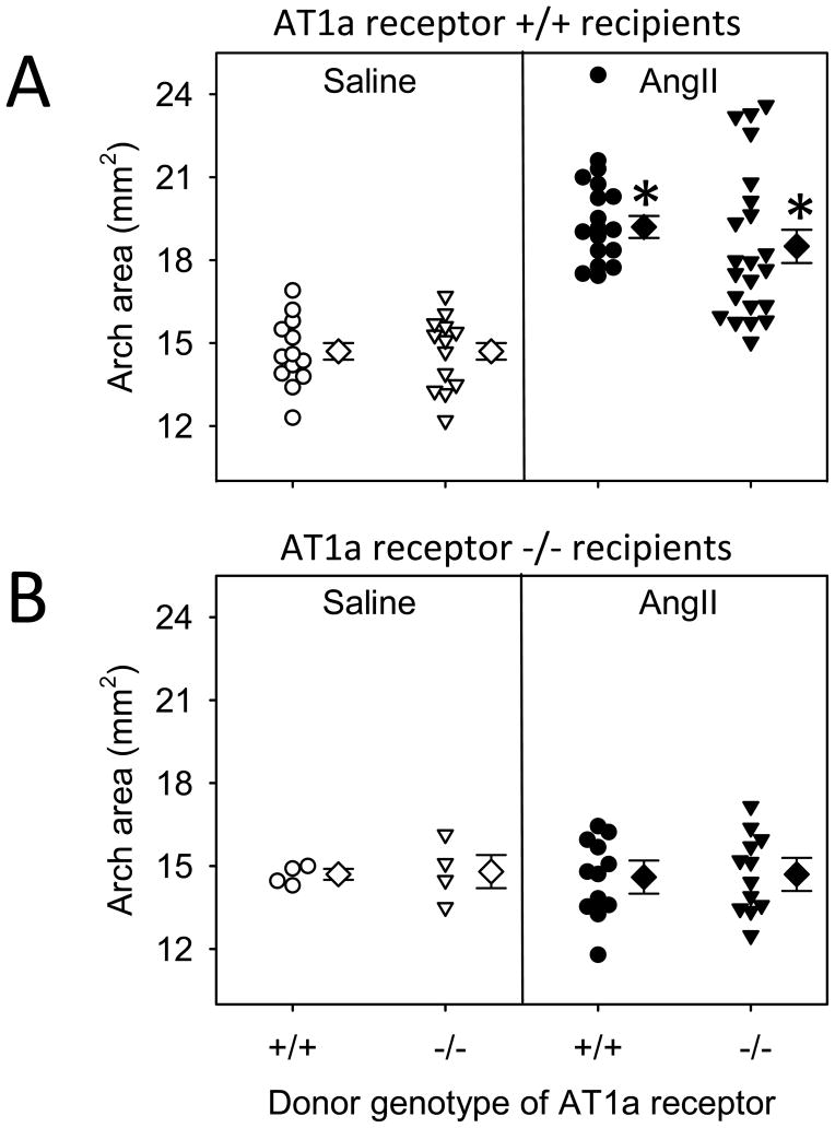
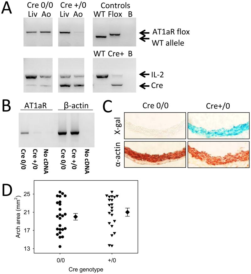
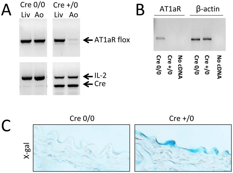
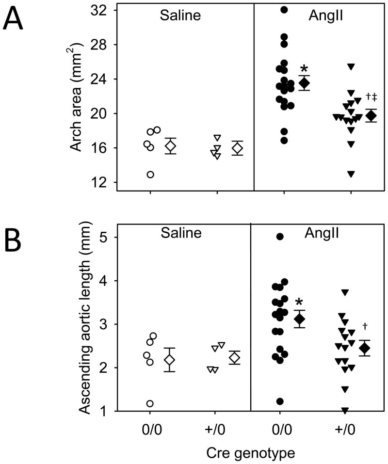
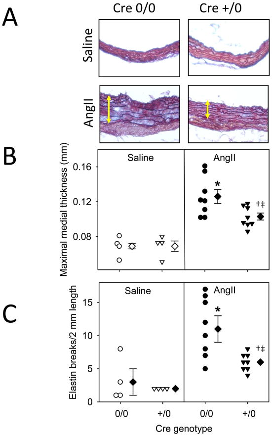
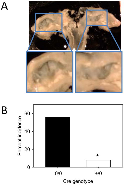
Methods and results: Male LDL receptor(-/-) mice were fed a saturated fat-enriched diet for 1 week before osmotic mini-pump implantation and infused with either saline or Ang II (1000 ng/kg per minute) for 28 days. Intimal surface areas of ascending aortas were measured to quantify ascending AAs. Whole body AT(1a) receptor deficiency ablated Ang II-induced ascending AAs (P<0.001). To determine the role of AT(1a) receptors on leukocytes, LDL receptor(-/-)×AT(1a) receptor(+/+) or AT(1a) receptor(-/-) mice were irradiated and repopulated with bone marrow-derived cells isolated from either AT(1a) receptor(+/+) or AT(1a) receptor(-/-) mice. Deficiency of AT(1a) receptors in bone marrow-derived cells had no effect on Ang II-induced ascending AAs. To determine the role of AT(1a) receptors on vascular wall cells, we developed AT(1a) receptor floxed mice with depletion on either smooth muscle or endothelial cells using Cre driven by either SM22 or Tek, respectively. AT(1a) receptor deletion in smooth muscle cells had no effect on ascending AAs. In contrast, endothelial-specific depletion attenuated this pathology.

Conclusions: Ang II infusion promotes aneurysms in the ascending aorta via stimulation of AT(1a) receptors that are expressed on endothelial cells.


Figures

Figure 1
Figure 1. Whole body deficiency of AT1a receptors ablated AngII-induced ascending AAs
Circles and triangles represent data from individual mice, diamonds are means, and bars are SEMs. * Denotes P<0.001 when comparing AngII versus saline infusion within +/+ group and when comparing +/+ versus −/− genotype in AngII infusion by two way ANOVA followed by the Holm-Sidak method.
Figure 2
Figure 2. Deficiency of AT1a receptors in bone marrow-derived cells did not affect AngII-induced ascending AAs
AT1a receptor +/+ (A) or −/− (B) recipients were irradiated and repopulated with bone marrow-derived cells harvested from either AT1a receptor +/+ or −/− mice. After repopulation, chimeric mice were infused with either saline or AngII. Circles and triangles represent individual mice, diamonds are means, and bars are SEMs. Statistical analyses were performed using two way ANOVA. There was no significant effect of genotype of the donor cells on aortic arch intimal areas.
Figure 3
Figure 3. AngII-induced aortic contractions were restricted to the infrarenal region
Regional contractility of rings from the (A) ascending, (B) thoracic, and (C) infrarenal aorta. Aortic rings were contracted during 5 minute incubations with potassium chloride (KCl, 80 mM), 5-HT (1 μM) and AngII (1 μM). Contractions are represented as a percentage of the maximal contraction achieved during incubation with elevated KCl (80 mM). Inverted arrows indicate the return to Krebs Henseleit solution containing no drug.
Figure 4
Figure 4. SMC deficiency of AT1a receptor had no effect on AngII-induced ascending AAs
Validation of AT1a receptor depletion in aortic smooth muscle cells (Ao) by PCR detection of (A) genomic DNA (compared to liver; Liv), and (B) mRNA. (C) ROSA26 Cre+/0 aortas were positive for β-galactosidase activity (blue color) in the same regions as positivity for alpha actin immunostaning. (D) Deficiency of AT1a receptors on SMCs had no significant effect on AngII-induced expansion of ascending aortas as defined by a Mann Whitney Rank Sum test. Data are from hemizygous Cre expressing mice (+/0) compared to Cre-negative littermates (0/0). Circles and triangles represent data from individual mice, diamonds are means, and bars are SEMs.
Figure 5
Figure 5. Validation of AT1a receptor depletion in endothelial cells
Validation was performed by PCR of: (A) genomic DNA and (B) mRNA by real time PCR with amplicons visualized on agarose gels. (C) ROSA26 Cre+/0 aortas were positive for β-galactosidase activity (blue color).
Figure 6
Figure 6. Endothelial cell-specific deficiency of AT1a receptors attenuated AngII-induced ascending AAs
(A) Intimal areas of ascending aortas. * Denotes P< 0.001 comparing saline versus AngII infusions in 0/0 genotype and comparing saline versus AngII infusions within genotypes; † denotes P=0.035 comparing saline versus AngII infusions within +/0 groups; ‡ denotes P=0.001 comparing 0/0 versus +/0 within AngII infusions by two way ANOVA. (B) Elongation measurement of outer curvature from aortic root to the brachiocephalic branch of the aorta. * Denotes P=0.016 comparing saline versus AngII infusions within 0/0; † denotes P=0.014 comparing 0/0 versus +/0 within AngII infusion.
Figure 7
Figure 7. Depletion of AT1a receptors in endothelium reduced medial thickness and elastin breaks
(A) A representative photomicrograph of saline and AngII infused AAs in Tek Cre 0/0 and Cre+/0 mice stained with Movat’s Pentachrome. (B) Medial thickness was measured from internal to external lamina. Circles and triangles represent data from individual mice, diamonds are means, and bars are SEMs. * Denotes P=<0.001 when comparing saline versus AngII infusion within 0/0; † denotes P=0.003 when comparing saline versus AngII infusion within +/0; ‡ denotes P=0.01 when comparing 0/0 versus +/0 within AngII infusion. (C) Elastin breaks were counted in each section. Circles and triangles represent data from individual mice, diamonds are means, and bars are SEMs. * Denotes P=<0.001 when comparing saline versus AngII infusion within 0/0; † denotes P=0.044 when comparing saline versus AngII infusion within +/0; ‡ denotes P=0.002 when comparing 0/0 versus +/0 within AngII infusions.
Figure 8
Figure 8. Endothelial cell-specific deficiency of AT1a receptors reduced the incidence of ascending aortic ulcers
(A) An example of an ulcerated ascending aorta. (B) The incidence of ulceration in ascending aortas of AngII infused groups. * Denotes P=0.004 by Fisher’s Exact test.

Comment in

  • Parsing aortic aneurysms: more surprises.
    Majesky MW, Dong XR, Hoglund VJ. Majesky MW, et al. Circ Res. 2011 Mar 4;108(5):528-30. doi: 10.1161/CIRCRESAHA.111.240861. Circ Res. 2011. PMID: 21372288 Free PMC article.

References

    1. Hiratzka LF, Bakris GL, Beckman JA, Bersin RM, Carr VF, Casey DE, Jr, Eagle KA, Hermann LK, Isselbacher EM, Kazerooni EA, Kouchoukos NT, Lytle BW, Milewicz DM, Reich DL, Sen S, Shinn JA, Svensson LG, Williams DM. 2010 ACCF/AHA/AATS/ACR/ASA/SCA/SCAI/SIR/STS/SVM guidelines for the diagnosis and management of patients with Thoracic Aortic Disease: a report of the American College of Cardiology Foundation/American Heart Association Task Force on Practice Guidelines, American Association for Thoracic Surgery, American College of Radiology, American Stroke Association, Society of Cardiovascular Anesthesiologists, Society for Cardiovascular Angiography and Interventions, Society of Interventional Radiology, Society of Thoracic Surgeons, and Society for Vascular Medicine. Circulation. 2010;121:e266–369. - PubMed
    1. Elefteriades JA, Farkas EA. Thoracic aortic aneurysm clinically pertinent controversies and uncertainties. J Am Coll Cardiol. 2010;55:841–857. - PubMed
    1. Brooke BS, Habashi JP, Judge DP, Patel N, Loeys B, Dietz HC., 3rd Angiotensin II blockade and aortic-root dilation in Marfan’s syndrome. N Engl J Med. 2008;358:2787–2795. - PMC - PubMed
    1. Pannu H, Tran-Fadulu V, Papke CL, Scherer S, Liu Y, Presley C, Guo D, Estrera AL, Safi HJ, Brasier AR, Vick GW, Marian AJ, Raman CS, Buja LM, Milewicz DM. MYH11 mutations result in a distinct vascular pathology driven by insulin-like growth factor 1 and angiotensin II. Hum Mol Genet. 2007;16:3453–3462. - PMC - PubMed
    1. Habashi JP, Judge DP, Holm TM, Cohn RD, Loeys BL, Cooper TK, Myers L, Klein EC, Liu G, Calvi C, Podowski M, Neptune ER, Halushka MK, Bedja D, Gabrielson K, Rifkin DB, Carta L, Ramirez F, Huso DL, Dietz HC. Losartan, an AT1 antagonist, prevents aortic aneurysm in a mouse model of Marfan syndrome. Science. 2006;312:117–121. - PMC - PubMed

Publication types

MeSH terms