. 2011 Apr;21(4):525-34.
doi: 10.1101/gr.114116.110. Epub 2011 Jan 20.

Large duplications at reciprocal translocation breakpoints that might be the counterpart of large deletions and could arise from stalled replication bubbles

Affiliations

Large duplications at reciprocal translocation breakpoints that might be the counterpart of large deletions and could arise from stalled replication bubbles

Karen D Howarth et al. Genome Res. 2011 Apr.

Abstract

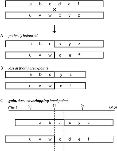
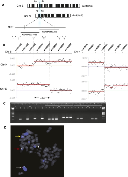
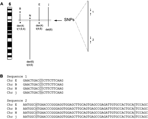
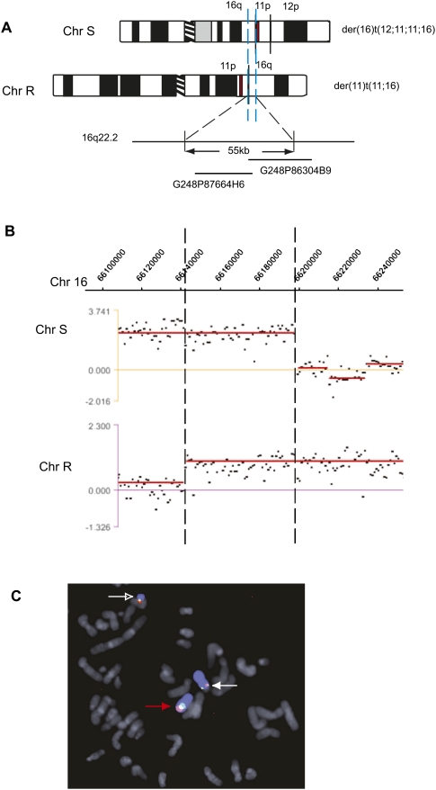
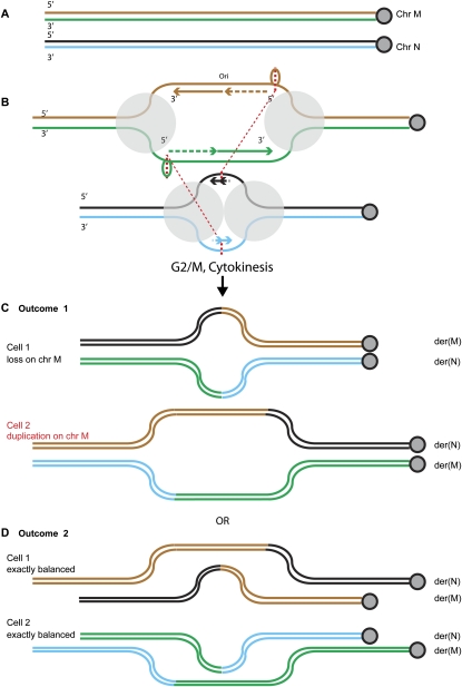
Reciprocal chromosome translocations are often not exactly reciprocal. Most familiar are deletions at the breakpoints, up to megabases in extent. We describe here the opposite phenomenon-duplication of tens or hundreds of kilobases at the breakpoint junction, so that the same sequence is present on both products of a translocation. When the products of the translocation are mapped on the genome, they overlap. We report several of these "overlapping-breakpoint" duplications in breast cancer cell lines HCC1187, HCC1806, and DU4475. These lines also had deletions and essentially balanced translocations. In HCC1187 and HCC1806, we identified five cases of duplication ranging between 46 kb and 200 kb, with the partner chromosome showing deletions between 29 bp and 31 Mb. DU4475 had a duplication of at least 200 kb. Breakpoints were mapped using array painting, i.e., hybridization of chromosomes isolated by flow cytometry to custom oligonucleotide microarrays. Duplications were verified by fluorescent in situ hybridization (FISH), PCR on isolated chromosomes, and cloning of breakpoints. We propose that these duplications are the counterpart of deletions and that they are produced at a replication bubble, comprising two replication forks with the duplicated sequence in between. Both copies of the duplicated sequence would go to one daughter cell, on different products of the translocation, while the other daughter cell would show deletion. These duplications may have been overlooked because they may be missed by FISH and array-CGH and may be interpreted as insertions by paired-end sequencing. Such duplications may therefore be quite frequent.


Figures

Figure 1.
Figure 1.
Breakpoint complexity at apparently balanced translocations. (A) Perfectly balanced rearrangement with no net gain or loss of material at the junction. (B) Deletion of material at the breakpoints of one or both translocation partners. (C) Duplication of material due to the same sequence being present on both products of a translocation—the breakpoints overlap on a genomic map (shown schematically for chromosome 1 as an example). Breakpoints are indicated with a dashed line, and the sequence in between is common to both translocation products.
Figure 2.
Figure 2.
HCC1806 t(4;6) duplication at a breakpoint. (A) Schematic representation of the products (Chr E and Chr N) of the reciprocal translocation between chromosomes 4 and 6 in HCC1806. The duplicated region of Chr 6 is shown between blue dashed lines, and the approximate location of fosmids used in FISH mapping is shown. The approximate location of chromosome 6 PCR primer pairs (1–6) is also shown. (B) Hybridization of Chr N and Chr E to a custom NimbleGen oligonucleotide array covering specified regions on chromosome 6 (left) and chromosome 4 (right). Breakpoints are indicated with a broken line. (C) Breakpoint mapping by PCR on flow-sorted chromosomes. PCR results with primer pairs 1–6 are shown. Lanes are labeled a (negative water control), b (HCC1806 genomic DNA), c (Chr N), and d (Chr E). (D) Breakpoint mapping by FISH using fosmids G248P81107D3 (shown in red) and G248P82010B5 (shown in green). Chromosome 6 is shown in blue. The der(4)t(1;6;4) is indicated with a red arrow (Chr B), the der(4)t(4;6) with a white arrow (Chr E), the der(6)t(4;6) with an open white arrow (Chr N), and the del (6) with a yellow arrow (Chr j). Other pieces of Chr 6 are the der(10)t(6;10) (Chr V), the der(14)t(6;14) (Chr Z), and the der(6)t(1;6) (Chr F) (Howarth et al. 2008).
Figure 3.
Figure 3.
Sequencing of SNPs at the duplicated chromosome 6 junction in HCC1806 reveals products of the reciprocal translocation between chromosomes 4 and 6 have the same haplotype. (A) An ideogram of chromosome 6 is shown, with abnormal chromosome 6 segments in HCC1806 indicated with black lines, labeled with their short chromosome names, chromosome B, etc. They are the der(4)t(1;6;4) (Chr B), the der(6)t(4;6) (Chr N), the der(4)t(4;6) (Chr E), and the del(6) (Chr j) (Howarth et al. 2008). Balanced breaks are indicated by *. The 46-kb region of chromosome 6 common to all chromosomes is highlighted with a gray box and shown expanded to the right. The approximate location of PCR primer pairs 1 and 2 is shown (giving PCR products of 250 bp and 350 bp, respectively; not to scale). (B) Examples of sequences from primer pairs 1 and 2 from the different flow-sorted chromosomes. SNPs are highlighted with gray boxes.
Figure 4.
Figure 4.
HCC1187 t(11;16) duplication at a breakpoint. (A) Schematic representation of the products of the reciprocal translocation between chromosomes 11 and 16 in HCC1187 (Chr S and Chr R). The duplicated region of Chr 16 is shown between blue dashed lines. The approximate location of fosmids used in FISH mapping is shown. A 1.3-kb duplicated piece of chromosome 11 is shown with a red bar. (B) Hybridization of Chr S and Chr R to a custom NimbleGen oligonucleotide array covering a specified region on chromosome 16. Breakpoints are indicated with a broken line. (C) Breakpoint mapping by FISH using fosmids G248P87664H6 (shown in red) and G248P86304B9 (shown in green). Chromosome 16 is shown in blue. Normal chromosome 16 is indicated with a red arrow, the der (16) with a white arrow (Chr S), and the der(11) with an open white arrow (Chr R) (confirming single-color FISH shown in Howarth et al. 2008).
Figure 5.
Figure 5.
Replication bubbles as a source of duplications and deletions at reciprocal translocation junctions. (A) Two normal chromosomes, M and N. Each DNA strand is shown as a thin line, centromere shown to the right. (B) Replication bubbles form, with the new strands shown. The polymerase complexes are shown as gray circles, the trombone loops at the lagging strand polymerase as ovals. For simplicity, the bubble on chromosome N is shown before significant synthesis has occurred, but it could be like the bubble on chromosome M, in which case, there are four possible outcomes. Breaks or template switching occurs at the thicker dotted red lines, perhaps at the single-stranded trombone or primer loops adjacent to the lagging strand polymerases, and reciprocal joining, i.e., translocation, occurs as shown by the thinner dotted lines, between them and breaks on chromosome N. Replication proceeds and the chromatids separate to daughter cells. According to which chromatids end up in the same cell, two outcomes are possible. (C) Outcome 1: Daughter cell 1 inherits a translocation with loss of the segment of chromosome M between the two breakpoints. Daughter cell 2 inherits two copies of this segment, to give a duplication at the breakpoint. (D) Outcome 2: Both cells receive exactly balanced products.
Figure 6.
Figure 6.
“Overlapping-breakpoint” duplications could be interpreted as an insertion by “paired-end read” methods. The duplication at the breakpoint on chromosome 6 of the t(4;6) in HCC1806 is shown in gray. Paired-end read fragments are shown as black bars joined with a dotted line. The same junction sequences (a, b, c, and d) would be generated by a reciprocal translocation with an “overlapping-breakpoint” duplication (above) or by an insertion of chromosome 6 (gray) into chromosome 4 (black) (below).

References

    1. Alsop AE, Taylor K, Zhang J, Gabra H, Paige AJ, Edwards PA 2008. Homozygous deletions may be markers of nearby heterozygous mutations: The complex deletion at FRA16D in the HCT116 colon cancer cell line removes exons of WWOX. Genes Chromosomes Cancer 47: 437–447 - PubMed
    1. Bignell GR, Santarius T, Pole JC, Butler AP, Perry J, Pleasance E, Greenman C, Menzies A, Taylor S, Edkins S, et al. 2007. Architectures of somatic genomic rearrangement in human cancer amplicons at sequence-level resolution. Genome Res 17: 1296–1303 - PMC - PubMed
    1. Bignell GR, Greenman CD, Davies H, Butler AP, Edkins S, Andrews JM, Buck G, Chen L, Beare D, Latimer C, et al. 2010. Signatures of mutation and selection in the cancer genome. Nature 463: 893–898 - PMC - PubMed
    1. Campbell PJ, Stephens PJ, Pleasance ED, O'Meara S, Li H, Santarius T, Stebbings LA, Leroy C, Edkins S, Hardy C, et al. 2008. Identification of somatically acquired rearrangements in cancer using genome-wide massively parallel paired-end sequencing. Nat Genet 40: 722–729 - PMC - PubMed
    1. Ciccone R, Giorda R, Gregato G, Guerrini R, Giglio S, Carrozzo R, Bonaglia MC, Priolo E, Lagana C, Tenconi R, et al. 2005. Reciprocal translocations: A trap for cytogenetists? Hum Genet 117: 571–582 - PubMed

Publication types

LinkOut - more resources