. 2011 Apr 1;286(13):10970-82.
doi: 10.1074/jbc.M110.166587. Epub 2011 Jan 20.

TDP-43 is a transcriptional repressor: the testis-specific mouse acrv1 gene is a TDP-43 target in vivo

Affiliations

TDP-43 is a transcriptional repressor: the testis-specific mouse acrv1 gene is a TDP-43 target in vivo

Avin S Lalmansingh et al. J Biol Chem. .

Abstract

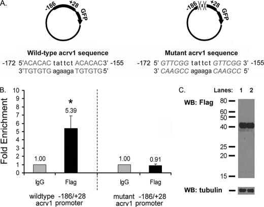
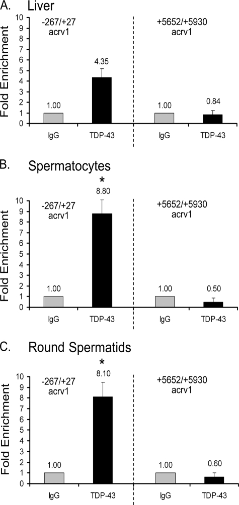
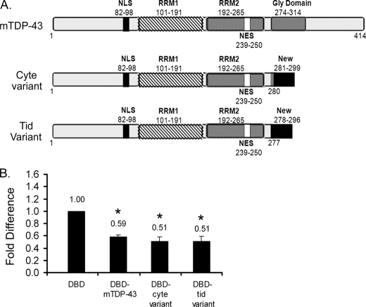
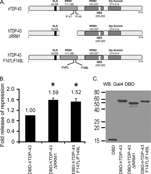
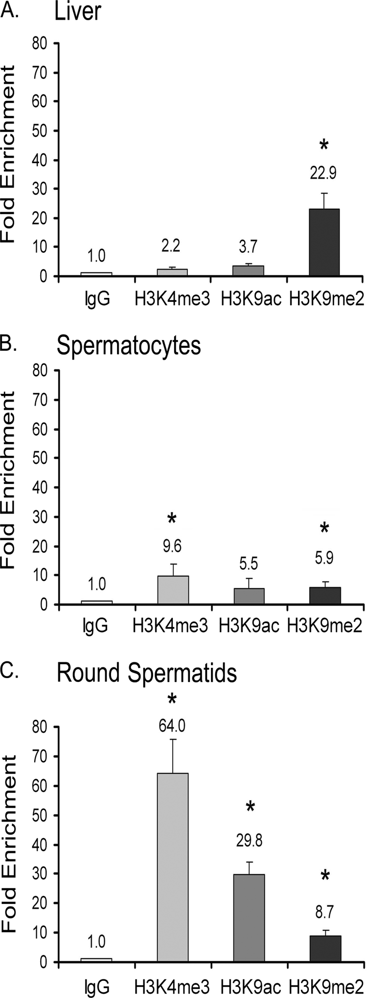
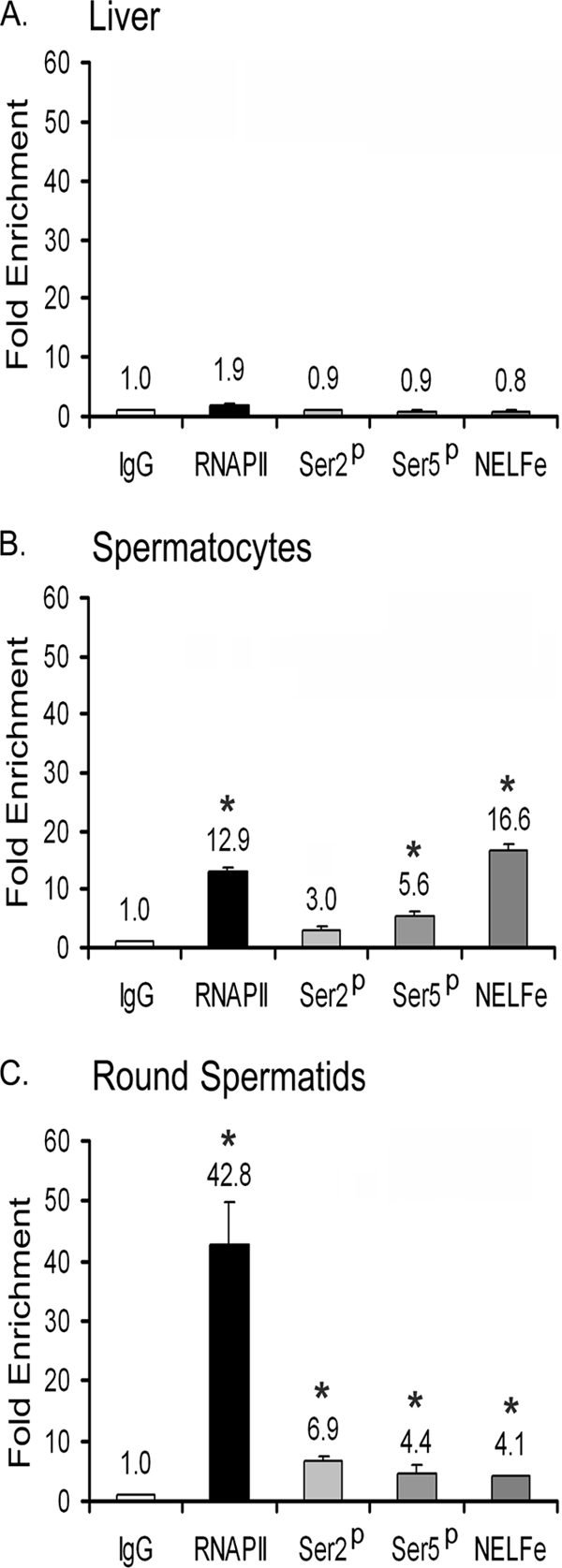
TDP-43 is an evolutionarily conserved ubiquitously expressed DNA/RNA-binding protein. Although recent studies have shown its association with a variety of neurodegenerative disorders, the function of TDP-43 remains poorly understood. Here we address TDP-43 function using spermatogenesis as a model system. We previously showed that TDP-43 binds to the testis-specific mouse acrv1 gene promoter in vitro via two GTGTGT-motifs and that mutation of these motifs led to premature transcription in spermatocytes of an otherwise round spermatid-specific promoter. The present study tested the hypothesis that TDP-43 represses acrv1 gene transcription in spermatocytes. Plasmid chromatin immunoprecipitation demonstrated that TDP-43 binds to the acrv1 promoter through GTGTGT motifs in vivo. Reporter gene assays showed that TDP-43 represses acrv1 core promoter-driven transcription via the N-terminal RRM1 domain in a histone deacetylase-independent manner. Consistent with repressor role, ChIP on physiologically isolated germ cells confirmed that TDP-43 occupies the endogenous acrv1 promoter in spermatocytes. Surprisingly, however, TDP-43 remains at the promoter in round spermatids, which express acrv1 mRNA. We show that RNA binding-defective TDP-43, but not splice variant isoforms, relieve repressor function. Transitioning from repressive to active histone marks has little effect on TDP-43 occupancy. Finally, we found that RNA polymerase II is recruited but paused at the acrv1 promoter in spermatocytes. Because mutation of TDP-43 sites caused premature transcription in spermatocytes in vivo, TDP-43 may be involved in pausing RNAPII at the acrv1 promoter in spermatocytes. Overall, our study shows that TDP-43 is a transcriptional repressor and that it regulates spatiotemporal expression of the acrv1 gene during spermatogenesis.


Figures

FIGURE 1.
FIGURE 1.
A–D, GAL4 recruitment strategy shows that full-length mouse TDP-43 represses transcription and that the N-terminal RRM domain is sufficient for transcriptional repression. A, in the schematic of the reporter gene used in this study five Gal4 binding sites were placed upstream of the −91/+28 acrv1 core promoter, which was fused to a luciferase reporter gene of the pGL3 basic vector to test TDP-43 repressor function. The GAL4 binding sites allow promoter recruitment of the GAL4 DBD-fusion proteins. B–D, transcriptional repressor function of mouse full-length TDP-43 (DBD-mTDP-43), various truncated forms (1–200, 1–262, 104–414, 191–414), and domains (RRM1, RRM2, RRM1 + 2, GLY) is shown. Schematics depicting mouse TDP-43 and major domains as DBD fusion proteins are shown to the left, and the reporter gene activities are shown to the right of each panel. One microgram of empty vector (DBD) or DBD-TDP-43 was co-transfected with 0.5 μg of reporter into GC-2 cells. 0.05 μg of Renilla Luciferase was used to normalize for transfection efficiency. Transcriptional output with DBD alone was used as the base line set as 1. FLAG-mTDP-43 is an untargeted TDP-43 version. p53AD corresponds to the activation domain of p53. Note: all DBD-TDP-43 clones showed nuclear localization (supplemental Fig. S1A) and migrated at the expected kDa sizes (supplemental Fig. S1B). Similar reporter assay results were obtained in the context of the c-fos reporter gene in HeLa cells (supplemental Fig. S2). NLS, nuclear localization signal; NES, nuclear export signal. E and F, HDAC inhibitors do not relieve TDP-43 mediated repression. TDP-43 mediated repression was evaluated in the presence of HDAC inhibitors sodium butyrate and Trichostatin A. Control DBD- and DBD-mTDP-43-transfected cells were treated with various concentrations of drug or vehicle (DMSO) for 24 h. Drug treatment effects are expressed as -fold difference of DMSO treatment. The inset shows dose-dependent hyperacetylation of core histones. HDAC inhibitors did not relieve TDP-43-mediated repression. NaB, sodium butyrate; TSA, Trichostatin A; Ac-H3, acetylated histone H3. All results shown (B–F) are the means ± S.E. for duplicate samples from four separate experiments. Asterisks represent significant difference (p < 0.05) compared with DBD. WB, Western blot.
FIGURE 2.
FIGURE 2.
Plasmid ChIP shows that TDP-43 binding to the acrv1 promoter is GTGTGT sequence-dependent. 3 μg of plasmid bearing either the wild-type −186/+28 mouse acrv1 promoter or a version bearing GTGTGT mutations were separately transfected into monkey kidney COS-7 cells. 3 μg of FLAG-TDP-43 was co-transfected. After 48 h, cells were harvested, and ChIP was performed with 5 μg of anti-FLAG antibody. A parallel ChIP with 5 μg of control IgG alone served as a negative control and provided base-line values. After ChIP, the −186/+27 region of the mouse acrv1 promoter was amplified using real-time quantitative PCR with plasmid-specific primers. PCR signal representing TDP-43 occupancy is plotted as -fold enrichment over IgG alone control. A, shown is a schematic of acrv1 promoter plasmids bearing wild-type and mutant promoter sequences. Nucleotide sequence of the region containing the TDP-43 recognition sequences (5′-GTGTGT) on the antisense strand is shown. B, results shown are the means ± S.E. for triplicate PCR samples from eight separate ChIP experiments. The asterisk represents significant difference (p < 0.05) compared with IgG as determined by one-way ANOVA followed by the Bonferroni post-hoc test. C, shown is a Western blot (WB) analysis of FLAG-TDP-43 expression in wild-type and mutant acrv1 plasmid transfected COS-7 cells.
FIGURE 3.
FIGURE 3.
TDP-43 is enriched at the endogenous acrv1 promoter in testicular germ cells. ChIP was performed on mouse liver cells, spermatocytes, and round spermatids using 5 μg of anti-TDP-43 polyclonal antibodies. A parallel ChIP of each cell type with 5 μg of IgG antibody alone served as a negative control and provided base-line values. The −267 to +27 region of the acrv1 proximal promoter, which includes the two TDP-43 binding sites at −172 and −160, was amplified by real-time PCR. Amplification of a downstream region of the acrv1 gene (+5652/+5930), which lacks GTGTGT sites, served as a negative control. PCR signal representing TDP-43 binding to the acrv1 gene is plotted as -fold increase over the IgG alone control. Results shown are the means ± S.E. for triplicate PCR samples from five separate ChIP experiments. The asterisks represent significant difference (p < 0.05) compared with IgG.
FIGURE 4.
FIGURE 4.
Splice variants of TDP-43 do not relieve repression. A, shown are schematics of TDP-43 splice variants cloned from mouse spermatocytes and round spermatids depicting that they lack the glycine-rich region but contain an additional 18 amino acids at the C-terminal end, not present in wild-type TDP-43. The spermatocyte splice variant contains three amino acids more than the round spermatid variant at position 278–280. B, shown is an evaluation of TDP-43 splice variants repressor function. 1 μg of empty vector (DBD) or DBD-TDP-43 was co-transfected with 0.5 μg of reporter into GC-2 cells. 0.05 μg of Renilla Luciferase was used to normalize for transfection efficiency. Transcriptional output with DBD alone was used as the base line set as 1. Note: DBD-TDP-43 variants show nuclear localization (supplemental Fig. S3B) and migrated at the expected molecular weight (supplemental Fig. S3C). The variants displayed similar repressor function in the context of the c-fos reporter gene in HeLa cells (supplemental Fig. S3D). Results shown are the means ± S.E. for duplicate samples from four separate experiments. NLS, nuclear localization signal; NES, nuclear export signal. The asterisks represent significant difference (p < 0.05) compared with DBD. Cyte, spermatocyte; Tid, round spermatid.
FIGURE 5.
FIGURE 5.
Deletion of RRM1 or mutation of Phe-147 and Phe-149 within RRM1 relieves TDP-43 repressor function. The −91/+28 acrv1 promoter luciferase reporter shown in Fig. 1A was used in this study. A, schematics of hTDP-43, hTDP-43 ΔRRM1, and hTDP-43 F147L/F149L clones are shown. NLS, nuclear localization signal; NES, nuclear export signal. B, transcriptional repressor function of DBD-hTDP-43, DBD-hTDP-43 ΔRRM1, and DBD-hTDP-43 F147L/F149L is shown. 1 μg of empty vector (DBD) or DBD-TDP-43 was co-transfected with 0.5 μg of reporter into GC-2 cells. 0.05 μg of Renilla Luciferase was used to normalize for transfection efficiency. Cells were harvested 48 h later, and luciferase activities were measured. The release of repression observed with ΔRRM1 and F147L/F149L mutants is expressed as -fold release over full-length hTDP-43 set as 1. Results shown are the means ± S.E. for duplicate samples from three separate experiments. Asterisk represent significant difference (p < 0.05) compared with DBD-hTDP-43. C, Western blot (WB) analysis showing expected kDa sizes of DBD-hTDP-43 clones using anti-DBD antibody (1:400) is shown. Note: all DBD-hTDP-43 clones show nuclear localization (supplemental Fig. S4).
FIGURE 6.
FIGURE 6.
Histone tail modifications associated with TDP-43 promoter occupancy in a physiological context. ChIP was performed on liver cells, spermatocytes, and round spermatids using 3 μg of IgG, H3K4me3, H3K9ac, and H3K9me2 antibodies. The −267 to +27 region of the acrv1 proximal promoter was amplified as before to determine factor binding. Amplification of a downstream region of the acrv1 gene (+5652/+5930) showed no factor binding (data not shown). A PCR signal representing chromatin marks associated with the acrv1 gene is plotted as -fold increase over the IgG alone control. Results shown are the means ± S.E. for triplicate PCR samples from four separate ChIP experiments. The asterisks represent significant difference (p < 0.05) compared with IgG.
FIGURE 7.
FIGURE 7.
Evidence for RNAPII pausing at the acrv1 promoter in spermatocytes and for its release in round spermatids. ChIP on liver cells, spermatocytes, and round spermatids was performed using 5 μg of IgG, total RNAPII-, RNAPII phosphoserine-2, phosphoserine 5-specific, and NELF-E antibodies. The −267 to +27 region of the acrv1 proximal promoter was amplified as before to determine factor binding. Amplification of a downstream region of the acrv1 gene (+5652/+5930) showed no factor binding (data not shown). PCR signal representing specific protein interactions with the acrv1 gene is plotted as -fold increase over the IgG alone control. Enrichment of phosphorylated Ser-5 of RNAPII and NELF-E at the promoter is indicative of paused RNAPII. Enrichment of phosphorylated Ser-2 mark and decrease of NELF-E occupancy in round spermatid is indicative of transcriptionally elongating RNAPII. Results shown are the means ± S.E. for triplicate PCR samples from 4 separate ChIP experiments. The asterisks represent significant difference (p < 0.05) compared with IgG. Ser2p, phosphoserine 2-specific RNAPII; Ser5p, phosphoserine 5-specific RNAPII.

Similar articles

Cited by

References

    1. Ou S. H., Wu F., Harrich D., García-Martínez L. F., Gaynor R. B. (1995) J. Virol. 69, 3584–3596 - PMC - PubMed
    1. Buratti E., Baralle F. E. (2008) Front. Biosci. 13, 867–878 - PubMed
    1. Neumann M., Sampathu D. M., Kwong L. K., Truax A. C., Micsenyi M. C., Chou T. T., Bruce J., Schuck T., Grossman M., Clark C. M., McCluskey L. F., Miller B. L., Masliah E., Mackenzie I. R., Feldman H., Feiden W., Kretzschmar H. A., Trojanowski J. Q., Lee V. M. (2006) Science 314, 130–133 - PubMed
    1. Chen-Plotkin A. S., Lee V. M., Trojanowski J. Q. (2010) Nat. Rev. Neurol. 6, 211–220 - PMC - PubMed
    1. Sephton C. F., Good S. K., Atkin S., Dewey C. M., Mayer P., 3rd, Herz J., Yu G. (2010) J. Biol. Chem. 285, 6826–6834 - PMC - PubMed

Publication types

MeSH terms

LinkOut - more resources