. 2011 Jul;18(7):1161-73.
doi: 10.1038/cdd.2010.184. Epub 2011 Jan 21.

Diacylglycerol kinase α regulates the formation and polarisation of mature multivesicular bodies involved in the secretion of Fas ligand-containing exosomes in T lymphocytes

Affiliations

Diacylglycerol kinase α regulates the formation and polarisation of mature multivesicular bodies involved in the secretion of Fas ligand-containing exosomes in T lymphocytes

R Alonso et al. Cell Death Differ. 2011 Jul.

Abstract

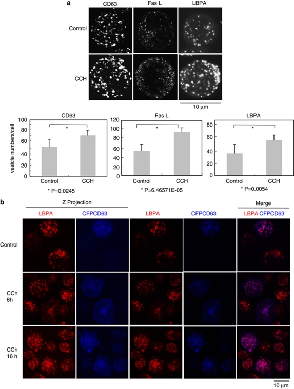
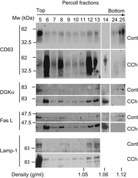
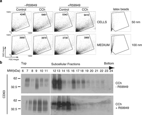
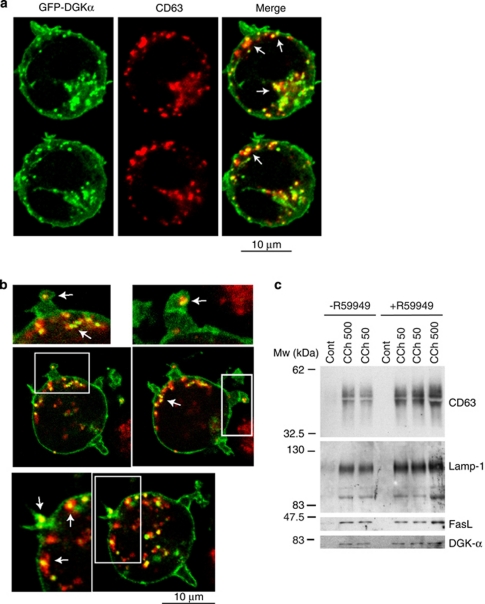
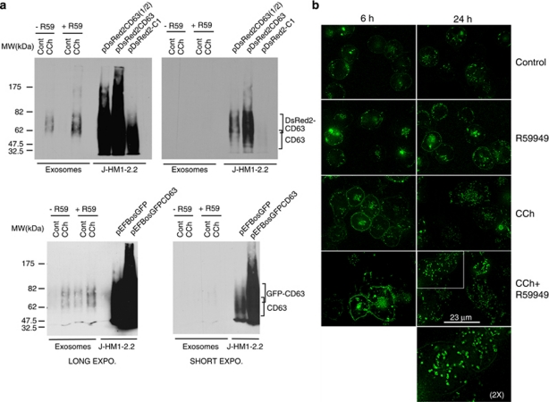
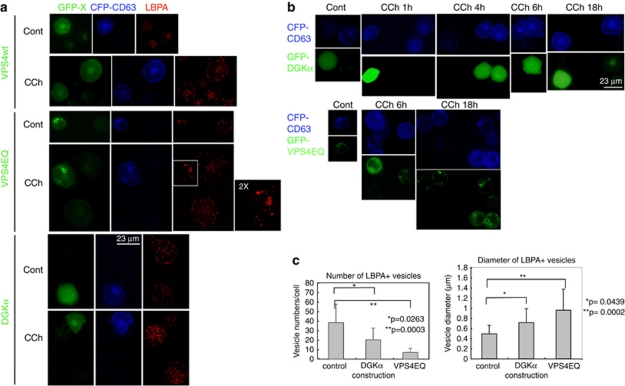
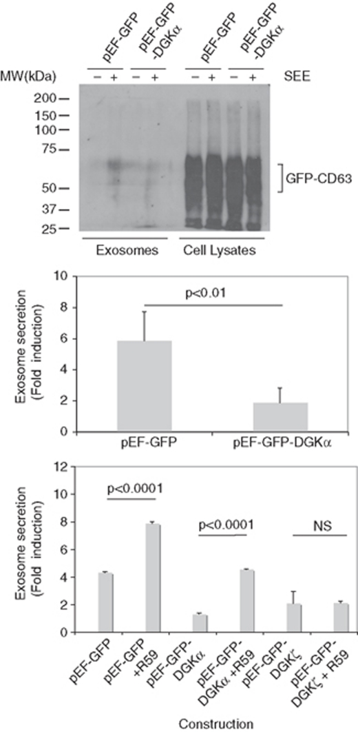
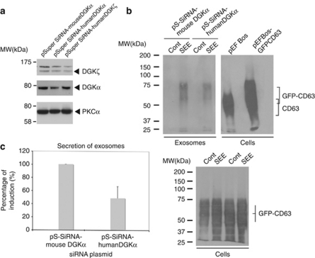
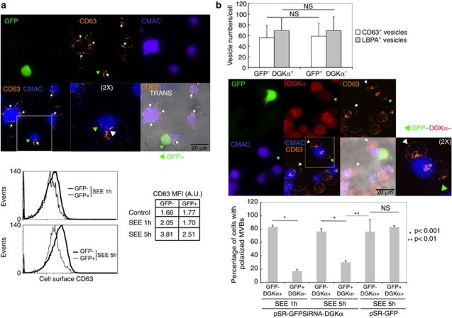
Multivesicular bodies (MVBs) are endocytic compartments that contain intraluminal vesicles formed by inward budding from the limiting membrane of endosomes. In T lymphocytes, these vesicles contain pro-apoptotic Fas ligand (FasL), which may be secreted as 'lethal exosomes' upon fusion of MVBs with the plasma membrane. Diacylglycerol kinase α (DGKα) regulate the secretion of exosomes, but it is unclear how this control is mediated. T-lymphocyte activation increases the number of MVBs that contain FasL. DGKα is recruited to MVBs and to exosomes in which it has a double function. DGKα kinase activity exerts a negative role in the formation of mature MVBs, as we demonstrate by the use of an inhibitor. Downmodulation of DGKα protein resulted in inhibition of both the polarisation of MVBs towards immune synapse and exosome secretion. The subcellular location of DGKα together with its complex role in the formation and polarised traffic of MVBs support the notion that DGKα is a key regulator of the polarised secretion of exosomes.


Figures

Figure 1
Figure 1
Cellular stimulation induces the formation of mature MVBs. (a) Upper panel, J-HM1-2.2 cells were stimulated with CCh for 6 h and then were imaged by confocal microscopy using antibodies specific for CD63, FasL and LBPA. Lamp-1 is mostly present on the limiting membrane of MVBs, whereas CD63, and particularly LBPA, are abundant in the ILVs, and therefore label mature MVBs. LBPA is a phospholipid that participates in the maturation of MVBs and constitutes a bonafide marker for ILVs of mature MVBs. The z-axis projections for each antigen corresponding to representative cells from three independent experiments are represented. Lower panel shows quantitative analysis of vesicles. Vesicle numbers were recorded from at least 20 cells per group, chosen randomly as indicated in Materials and Methods. Results represent average number of vesicles/cell±S.D. of three independent experiments. (b) J-HM1-2.2 cells, transfected with CFP-CD63, were stimulated with CCh (for 6 and 16 h), and mature MVBs were visualised with anti-LBPA antibody (red) as indicated in Materials and Methods. Cells were imaged by confocal microscopy and representative (n=3 independent experiments), single optical sections (0.4 μm thick) and merged images (coincident labelling appearing pink) are shown in the right side. In the left side, z-axis projection images of LBPA and CFP-CD63 are shown
Figure 2
Figure 2
Cellular stimulation induces the relocalisation of FasL and DGKα to subcellular fractions containing MVBs. Cellular fractionation by density gradient of the homogenates from equal numbers of J-HM1-2.2 cells, stimulated or not stimulated with CCh (6 h), was performed as indicated in Materials and Methods, and the Percoll fractions were analysed for CD63, DGKα and FasL by WB. The blot was reprobed with anti Lamp-1 antibody as a loading control. Data are representative of the results obtained in three different experiments
Figure 3
Figure 3
Inhibition of DGKα kinase activity increases the number of MVBs and the secretion of exosomes. (a) The secretion of exosomes was induced by treatment of J-HM1-2.2 cells with CCh during 10 h, preincubated or not with R59949 (10 μM). In the upper row, the dot plots correspond to the events recorded – the number of events is included inside each plot – in the cell culture supernatants from cells treated as indicated, whereas the lower row plots register the events from complete medium treated with different reagents but in the absence of cells. The lower row dot plots registered a constant number of events; thus events above this background should be considered as specific, cell-produced vesicles. In the right-hand side, dot plots corresponding to 50 and 100 nm latex beads, analysed in parallel, are included as a reference. (b) WB with anti-CD63 of Percoll density gradient fractions from cells stimulated with CCh for 6 h, pretreated or not with R59949. Data are representative of the results obtained in three different experiments. The shift in the distribution of CD63 on the density gradient is compatible with changes in the lipid composition and the maturation of MVBs
Figure 4
Figure 4
DGKα is associated to CD63+ vesicles and exosomes. (a) Cells expressing GFP-DGKα were stimulated with CCh for 6 h in the presence of R59949, stained with anti-CD63 (red), and cells were imaged by confocal microscopy; several representative, optical sections (0.4 μm thick) are shown. The right-hand panels show the merged images, with coincident labelling appearing yellow. (b) Digital zoom ( × 1.67) of some images from one experiment similar to that described in panel a are shown. (c) Cells were stimulated or not stimulated with CCh (50 and 500 μM) for 12 h in the presence or absence of R59949 (10 μM), and the secreted exosomes were analysed by WB for the presence of several exosome markers and endogenous DGKα. Results are representative of the data obtained in three different experiments
Figure 5
Figure 5
Expression of the reporters for MVBs/exosomes in living cells. (a) J-HM1-2.2 cells expressing DsRed2-CD63 (upper panels) or GFP-CD63 (lower panels) were stimulated with CCh for 10 h in the presence or the absence of R59949 inhibitor (R59, 10 μM), and the isolated exosomes were analysed by WB with anti-CD63 to detect the chimerical CD63 molecules. Different exposures of the same blot are shown to visualise both the chimeras and the endogenous CD63. In the right-side lanes of each blot, lysates from cells expressing or not expressing the CD63 chimeras were run as a reference. (b) Cells expressing GFP-CD63 were stimulated or not expressing with CCh for 6 and 24 h in the presence or the absence of R59949 to visualise the formation and fate of MVBs in living cells. The CCh- and R59949-induced increase in GFP-CD63 at the plasma membrane was probably because of the fusion of the limiting membrane of GFP-CD63+ MVBs (see also Supplementary Videos 3 and 4). The epifluorescence images were improved by image deconvolution as indicated in Materials and Methods, and are representative of the results obtained of more than 50 cells recorded per treatment in four independent experiments. The inset shows a × 2 digital zoom of the indicated area
Figure 6
Figure 6
Expression of GFP-DGKα affects the formation of mature MVBs and the distribution of LBPA+ vesicles, but does not inhibit the accumulation of CD63 at the plasma membrane. Jurkat cells expressing GFP-VPSwt, GFP-VPS4EQ mutant or GFP-DGKα, together with CFP-CD63, were stimulated with CCh to induce the formation and traffic of MVBs, and LBPA was stained as indicated in Materials and Methods. The endosomal sorting complex required for transport (ESCRT) machinery is involved in the formation of the intraluminal vesicles of MVBs, and the AAA-ATPase vacuolar protein-sorting VPS4 regulates the recycling of the ESCRT complexes. The expression of a VPS4 ATPase-defective mutant, VPS4EQ, inhibits the inward budding at the limiting membrane of MVBs and the subsequent formation of ILVs., These effects result in the formation of immature, large endosomes,, and thus the expression of VPS4EQ mutant constitutes an appropriate control for aberrant MVBs maturation. (a) Epifluorescence microscopy images (GFP-X, CFP-CD63 and LBPA, respectively) of control and CCh-stimulated (8 h) cells. In GFP-VPS4EQ-expressing cells, the dispersion of LBPA+ granules that was induced by CCh in GFP-VPS4EQ cells or in cells expressing GFP-VPS4wt was inhibited, and the large LBPA+ structures accumulated in the perinuclear area. (b) Cells expressing GFP-DGKα (upper rows) or GFP-VPS4EQ (lower rows), together with CFP-CD63 were stimulated with CCh for the indicated times to visualise the accumulation of CFP-CD63 at the plasma membrane by fluorescence microscopy. CD63 cell surface labelling is a consequence of the transport, docking and fusion of MVBs with the plasma membrane (see also Figure 5b, Supplementary Videos 3 and 4). (c) The average number per cell and the mean diameter (±S.D.) of LBPA+ vesicles were measured in cells from three independent experiments similar to that described in panel a (in a total of 11 control, untransfected cells; 8 GFP-DGKα+ cells; and 19 GFP-VPS4EQ+ cells) after 6 h of CCh stimulation. In a significant fraction (40–50%, n=20 cells) of GFP-DGKα-expressing cells, the number of LBPA+ vesicles was lower to that found in GFP-DGKα cells stimulated with CCh (Supplementary Figure S5, panel A, third row). In approximately 20% of GFP-DGKα+ cells, the LBPA+ structures were condensed in some areas but not dispersed throughout the cytosol (Supplementary Figure S5, panel A, lower row), as observed in GFP-VPS4EQ+ cells (Supplementary Figure S5, panel A, second row). See also Supplementary Figure S5, panel C
Figure 7
Figure 7
Modulation of DGKα pathway controls the polarised secretion of exosomes. Jurkat cells co-expressing GFP-DGKα or GFP, together with GFP-CD63, were mixed with Raji cells, pulsed or not with SEE, to induce the formation of synapse and the secretion of exosomes. Upper panel: the amount of exosomes produced by cells expressing similar amounts of GFP-CD63 (cell lysates, right side) at the end of the culture period was measured by WB with an antibody against CD63. Middle panel: quantification of the secretion of exosomes in cells expressing GFP-DGKα in comparison with cells expressing GFP. Results summarise the data obtained from WB in four independent experiments and are represented as average (±S.D.) fold induction of exosome secretion. Lower panel: the effect on exosome secretion of the pretreatment with R59949 (10 μM) of cells expressing GFP (control), GFP-DGKα or GFP-DGKζ was measured as indicated in the middle panel (n=3 independent experiments)
Figure 8
Figure 8
Interference of DGKα inhibits polarised exosome secretion. Jurkat cells were co-transfected with interference plasmids for human DGKα, human DGKζ, or an irrelevant, mouse DGKα, together with reporter GFP-CD63, and were stimulated with Raji cells, pulsed or not (control) with SEE, to induce secretion of exosomes. Transfection efficiency of Jurkat cells in these experiments was around 50%, as assessed by flow cytometry. (a) WB of Jurkat cells transfected with the interference plasmids before stimulation with Raji cells, showing the specificity of the interference with specific antibodies against DGKα and DGKζ. (b) WB of the exosomes (upper panel) isolated from the cell culture supernatants of the interfered cells after synapse formation (12 h). In the right side, extracts of cells transfected with an empty vector and a GFP-CD63 vector were analysed as reference. The WB of the cells (lower panel) recovered after the culture period was performed in parallel to normalise by viable cell number at the end of the culture period and by transfection efficiency of the GFP-CD63 reporter. (c) Summary of the results from four independent experiments similar to the one shown in panel b; results are represented as average (±S.D.) percent of SEE induction of exosome secretion
Figure 9
Figure 9
Interference of DGKα affects polarisation and degranulation of MVBs at the immune synapse. Jurkat cells transfected with a GFP-containing, bicistronic interference plasmid for human DGKα were stimulated for 1 and 5 h with Raji cells previously pulsed with SEE and labelled with CMAC (blue), to induce synapse formation and the polarised traffic of MVBs. (a) Upper panel: Degranulation of MVBs at the synapse after 5 h was assessed by cell surface staining of endogenous CD63 (orange-red) and fluorescence microscopy. White arrowheads label the synaptic contact areas made by GFP Jurkat cells, whereas green arrowhead labels the synapse corresponding to a GFP+ Jurkat cell. Images are representative of the results obtained in four different experiments. Lower panel: degranulation induced by SEE was measured after 1 and 5 h, by analysing cell surface staining of CD63 in both GFP+ (DGKα) and GFP (DGKα+) Jurkat cells by flow cytometry. The FACS profiles corresponding to the staining of Jurkat cells challenged with Raji without SEE are not shown for clarity, but the corresponding mean fluorescence intensity data are included (control). (b) Upper panel: quantification of LBPA+ and CD63+ intracellular vesicles in Jurkat cells forming synapse with SEE-pulsed Raji cells during 5 h. Middle panel: polarisation of MVBs towards the synapse after 5 h was assessed by intracellular labelling of endogenous CD63 (orange-red). Interference with DGKα expression was tested by intracellular staining of endogenous DGKα (red). White arrowheads label synapses made by GFP DGKα+ Jurkat cells, whereas green arrowhead labels the synapse corresponding to a GFP+ DGKα Jurkat cell. Images are representative of the results obtained in four different experiments. The inset shows a × 2 digital zoom of the indicated areas. Lower panel: the graph summarises the results of four independent experiments (at least 40 cells per condition were analysed) similar to the one described in the middle panel, and compares the percentage of cells with polarised MVBs after 1 and 5 h of stimulation, both in DGKα+ cells and in DGKα cells. Cells with polarised MVBs were defined as those cells on which the majority of their MVBs were located at a distance of the synapse lower than one quarter of the diameter of the cell. The interference on DGKα expression does not affect the interaction among T lymphocytes and the SEE-presenting cells, as the frequency of formation of Raji/Jurkat conjugates was not affected by DGKα interference (not shown)

References

    1. Stoorvogel W, Kleijmeer MJ, Geuze HJ, Raposo G. The biogenesis and functions of exosomes. Traffic. 2002;3:321–330. - PubMed
    1. Bossi G, Griffiths GM. Degranulation plays an essential part in regulating cell surface expression of Fas ligand in T cells and natural killer cells. Nat Med. 1999;5:90–96. - PubMed
    1. Nagata S. Apoptosis by death factor. Cell. 1997;88:355–365. - PubMed
    1. Zuccato E, Blott EJ, Holt O, Sigismund S, Shaw M, Bossi G, et al. Sorting of Fas ligand to secretory lysosomes is regulated by mono-ubiquitylation and phosphorylation. J Cell Sci. 2007;120:191–199. - PubMed
    1. Monleon I, Martinez-Lorenzo MJ, Monteagudo L, Lasierra P, Taules M, Iturralde M, et al. Differential secretion of Fas ligand- or APO2 ligand/TNF-related apoptosis-inducing ligand-carrying microvesicles during activation-induced death of human T cells. J Immunol. 2001;167:6736–6744. - PubMed

Publication types

MeSH terms