Vardenafil Enhances Oxytocin Expression in the Paraventricular Nucleus without Sexual Stimulation
- PMID: 21253331
- PMCID: PMC3021811
- DOI: 10.5213/inj.2010.14.4.213
Vardenafil Enhances Oxytocin Expression in the Paraventricular Nucleus without Sexual Stimulation
Abstract
Purpose: Oxytocin is associated with the ability to form normal social attachments. c-Fos is an immediate early gene whose expression is used as a marker for stimulus-induced changes in neurons. The effect of phosphodiesterase-5 (PDE-5) inhibitors on oxytocin activation in the brain without sexual stimuli has not yet been reported. In the present study, we investigated the effects of vardenafil on oxytocin and c-Fos expression in the paraventricular nucleus (PVN) of conscious rats.
Methods: Male Sprague-Dawley rats weighing 300±10 g were divided into 6 groups (n=5 in each group): the control group, the 1-day-0.5 mg/kg, the 1-day-1 mg/kg, the 1-day-2 mg/kg, the 3-day-1 mg/kg, and the 7-day-1 mg/kg vardenafil administration group. The experiment was conducted without sexual stimulation. Vardenafil was orally administered. The animals in the control group received an equivalent amount of distilled water orally. The expression of oxytocin and c-Fos in the PVN was detected by immunohistochemistry.
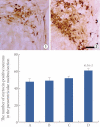
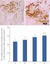
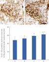
Results: Oxytocin expression in the PVN was increased by 1 day administration of 2 mg/kg vardenafil, and this effect of vardenafil appeared in a duration-dependent manner. c-Fos in the oxytocin neurons of the PVN was increased by 1 day administration of 2 mg/kg vardenafil, and this effect of vardenafil also appeared in a duration-dependent manner. These results showed that vardenafil augments the expression of oxytocin with activation of oxytocin neurons in the PVN.
Conclusions: In this study, we showed that the PDE-5 inhibitor, vardenafil directly enhances oxytocin expression and also activates oxytocin neurons in the PVN, which indicates that vardenafil may exert positive effects on affiliation behavior and social interaction.
Keywords: Oxytocin; Paraventricular nucleus; Rats; Vardenafil; c-Fos.
Conflict of interest statement
No potential conflict of interest relevant to this article was reported.
Figures


Similar articles
-
Effect of vardenafil on nitric oxide synthase expression in the paraventricular nucleus of rats without sexual stimulation.Andrologia. 2012 May;44 Suppl 1:56-67. doi: 10.1111/j.1439-0272.2010.01138.x. Epub 2011 Sep 24. Andrologia. 2012. PMID: 21950284
-
Central administration of cocaine-amphetamine-regulated transcript activates hypothalamic neuroendocrine neurons in the rat.Endocrinology. 2000 Feb;141(2):794-801. doi: 10.1210/endo.141.2.7295. Endocrinology. 2000. PMID: 10650962
-
Central administration of glucagon-like peptide-1 activates hypothalamic neuroendocrine neurons in the rat.Endocrinology. 1997 Oct;138(10):4445-55. doi: 10.1210/endo.138.10.5270. Endocrinology. 1997. PMID: 9322962
-
Intravenous CDP-choline activates neurons in supraoptic and paraventricular nuclei and induces hormone secretion.Brain Res Bull. 2012 Feb 10;87(2-3):286-94. doi: 10.1016/j.brainresbull.2011.11.013. Epub 2011 Nov 27. Brain Res Bull. 2012. PMID: 22138197
-
Different signaling molecules responsible for IL-1beta-induced oxytocinergic and vasopressinergic neuron activation in the hypothalamic paraventricular nucleus of the rat.Neurochem Int. 2006 Mar;48(4):312-7. doi: 10.1016/j.neuint.2005.11.007. Epub 2006 Jan 18. Neurochem Int. 2006. PMID: 16386822
Cited by
-
Oxytocin: a therapeutic target for mental disorders.J Physiol Sci. 2012 Nov;62(6):441-4. doi: 10.1007/s12576-012-0232-9. Epub 2012 Sep 25. J Physiol Sci. 2012. PMID: 23007624 Free PMC article. Review.
References
-
- Pedersen CA. Oxytocin control of maternal behavior. Regulation by sex steroids and offspring stimuli. Ann N Y Acad Sci. 1997;807:126–145. - PubMed
-
- Carter CS. Neuroendocrine perspectives on social attachment and love. Psychoneuroendocrinology. 1998;23:779–818. - PubMed
-
- Uvnäs-Moberg K. Oxytocin may mediate the benefits of positive social interaction and emotions. Psychoneuroendocrinology. 1998;23:819–835. - PubMed
-
- Landgraf R, Neumann ID. Vasopressin and oxytocin release within the brain: a dynamic concept of multiple and variable modes of neuropeptide communication. Front Neuroendocrinol. 2004;25:150–176. - PubMed
-
- Huber D, Veinante P, Stoop R. Vasopressin and oxytocin excite distinct neuronal populations in the central amygdala. Science. 2005;308:245–248. - PubMed
LinkOut - more resources
Full Text Sources

