

Left atrial mass with stalk: thrombus or myxoma?
- PMID: 21253367
- PMCID: PMC3021896
- DOI: 10.4250/jcu.2010.18.4.154
Left atrial mass with stalk: thrombus or myxoma?
Abstract
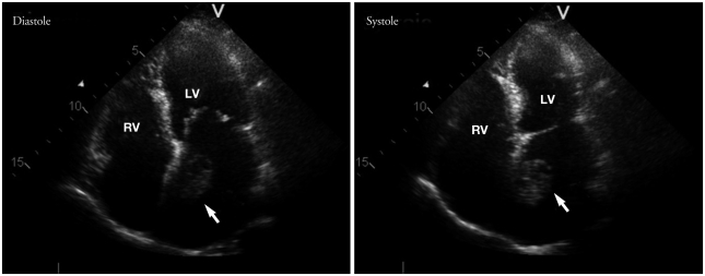
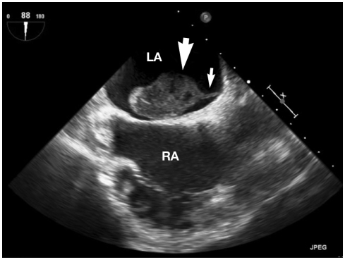
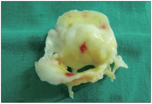
A 63-year-old female was presented to emergency room with an abdominal pain. The patient had moderate mitral valve stenosis and atrial fibrillation. Abdominal computed tomography revealed right renal infarction. Transthoracic echocardiography showed a large mobile mass in the left atrium. Transesophageal two-and three-dimensional echocardiography showed a large mobile ovoid mass with a narrow stalk attached to the left atrial septum. It was thought to be a myxoma rather than thrombus. Anticoagulation with heparin was continued. When the operation was performed, there was no mass in the left atrium. It must be a thrombus and melt away.
Keywords: Atrial fibrillation; Left atrium; Myxoma; Stalk; Thrombus.
Figures

References
-
- Alam M, Sun I. Transesophageal echocardiographic evaluation of left atrial mass lesions. J Am Soc Echocardiogr. 1991;4:323–330. - PubMed
-
- Feinglass NG, Reeder GS, Finck SJ, Shine TS, Maniu CV. Myxoma of the left atrial appendage mimicking thrombus during aortic valve replacement. J Am Soc Echocardiogr. 1998;11:677–679. - PubMed
-
- Araoz PA, Mulvagh SL, Tazelaar HD, Julsrud PR, Breen JF. CT and MR imaging of benign primary cardiac neoplasms with echocardiographic correlation. Radiographics. 2000;20:1303–1319. - PubMed
-
- Fang YM, Dean R, Figueroa R. Right atrial myxoma mimicking an atrial thrombus in the third trimester of pregnancy. J Matern Fetal Neonatal Med. 2007;20:77–78. - PubMed
-
- Feinglass NG, Reeder GS, Finck SJ, Shine TS, Maniu CV. Myxoma of the left atrial appendage mimicking thrombus during aortic valve replacement. J Am Soc Echocardiogr. 1998;11:677–679. - PubMed
Publication types
LinkOut - more resources
Full Text Sources
