

Crystalline ultrastructures, inflammatory elements, and neoangiogenesis are present in inconspicuous aortic valve tissue
- PMID: 21253468
- PMCID: PMC3022178
- DOI: 10.4061/2010/685926
Crystalline ultrastructures, inflammatory elements, and neoangiogenesis are present in inconspicuous aortic valve tissue
Abstract
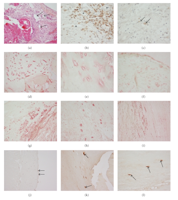
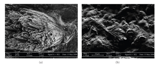
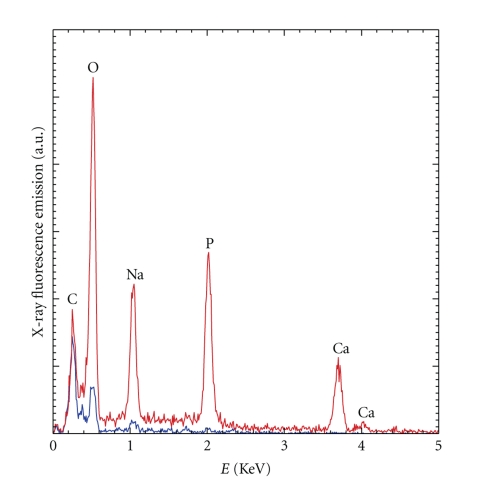
Morbidity from calcific aortic valve disease (CAVD) is increasing. Recent studies suggest early reversible changes involving inflammation and neoangiogenesis. We hypothesized that microcalcifications, chemokines, and growth factors are present in unaffected regions of calcific aortic valves. We studied aortic valves from 4 patients with CAVD and from 1 control, using immunohistochemistry, scanning electron microscopy, and infrared spectrography. We revealed clusters of capillary neovessels in calcified (ECC), to a lesser extent in noncalcified (ECN) areas. Endothelial cells proved constant expression of SDF-1 in ECC, ECN, and endothelial cells from valvular surface (ECS). Its receptor CXCR4 was expressed in ECC. IL-6 expression correlated with CXCR4 staining and presence of lymphocytes. VEGF was expressed by ECS, its receptor by ECC and ECN. Crystalline ultrastructures were found on the surface of histologically noncalcified areas (HNCAs), spectrography revealed calcium hydroxylapatite. Our results demonstrate that crystalline ultrastructures are present in HNCAs, undergoing neoangiogenesis in an inflammatory context. These alterations could be an early witness of disease and an opening to therapy.
Figures
Similar articles
-
CD39 and CD73 in the aortic valve-biochemical and immunohistochemical analysis in valve cell populations and its changes in valve mineralization.Cardiovasc Pathol. 2018 Sep-Oct;36:53-63. doi: 10.1016/j.carpath.2018.05.008. Epub 2018 Jun 7. Cardiovasc Pathol. 2018. PMID: 30056298
-
Histopathological assessment of calcification and inflammation of calcific aortic valves from patients with and without diabetes mellitus.Histol Histopathol. 2017 Mar;32(3):293-306. doi: 10.14670/HH-11-797. Epub 2016 Jun 29. Histol Histopathol. 2017. PMID: 27353274 Free PMC article.
-
Amyloid substance within stenotic aortic valves promotes mineralization.Histopathology. 2012 Oct;61(4):610-9. doi: 10.1111/j.1365-2559.2012.04265.x. Histopathology. 2012. PMID: 22642224
-
Calcific Aortic Valve Disease-Natural History and Future Therapeutic Strategies.Front Pharmacol. 2020 May 13;11:685. doi: 10.3389/fphar.2020.00685. eCollection 2020. Front Pharmacol. 2020. PMID: 32477143 Free PMC article. Review.
-
Calcific aortic valve disease: is it another face of atherosclerosis?Indian Heart J. 2015 Sep-Oct;67(5):503-6. doi: 10.1016/j.ihj.2015.07.033. Epub 2015 Aug 21. Indian Heart J. 2015. PMID: 26432749 Free PMC article. Review.
Cited by
-
Extracellular Matrix in Calcific Aortic Valve Disease: Architecture, Dynamic and Perspectives.Int J Mol Sci. 2021 Jan 18;22(2):913. doi: 10.3390/ijms22020913. Int J Mol Sci. 2021. PMID: 33477599 Free PMC article. Review.
-
Prostatic stones: evidence of a specific chemistry related to infection and presence of bacterial imprints.PLoS One. 2012;7(12):e51691. doi: 10.1371/journal.pone.0051691. Epub 2012 Dec 13. PLoS One. 2012. PMID: 23272143 Free PMC article.
-
Role of endothelial CXCR4 in the development of aortic valve stenosis.Front Cardiovasc Med. 2022 Sep 6;9:971321. doi: 10.3389/fcvm.2022.971321. eCollection 2022. Front Cardiovasc Med. 2022. PMID: 36148060 Free PMC article.
-
M1/M2 macrophages and associated mechanisms in congenital bicuspid aortic valve stenosis.Exp Ther Med. 2014 Apr;7(4):935-940. doi: 10.3892/etm.2014.1529. Epub 2014 Feb 10. Exp Ther Med. 2014. PMID: 24669254 Free PMC article.
-
Characterization of porcine aortic valvular interstitial cell 'calcified' nodules.PLoS One. 2012;7(10):e48154. doi: 10.1371/journal.pone.0048154. Epub 2012 Oct 26. PLoS One. 2012. PMID: 23110195 Free PMC article.
References
-
- O’Brien KD. Pathogenesis of calcific aortic valve disease: a disease process comes of age (and a good deal more) Arteriosclerosis, Thrombosis, and Vascular Biology. 2006;26(8):1721–1728. - PubMed
-
- Li YB, Hu CL, Liu J, Tang QIZ, Huang CX. Calcific aortic valve disease should not be considered as a degenerative disease anymore. Medical Hypotheses. 2007;68(6):1233–1235. - PubMed
-
- Rosenhek R, Rader F, Loho N, et al. Statins but not angiotensin-converting enzyme inhibitors delay progression of aortic stenosis. Circulation. 2004;110(10):1291–1295. - PubMed
-
- Mohler ER, Gannon F, Reynolds C, Zimmerman R, Keane MG, Kaplan FS. Bone formation and inflammation in cardiac valves. Circulation. 2001;103(11):1522–1528. - PubMed
LinkOut - more resources
Full Text Sources