

Alzheimer's disease and glaucoma: imaging the biomarkers of neurodegenerative disease
- PMID: 21253485
- PMCID: PMC3022203
- DOI: 10.4061/2010/793931
Alzheimer's disease and glaucoma: imaging the biomarkers of neurodegenerative disease
Abstract
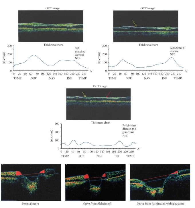
Imaging through the visual system in Alzheimer's disease, with the technology currently in widespread use for the diagnosis and management of eye disease such as glaucoma and macular degeneration, is proving to be promising. In vivo cross-section imaging during an annual comprehensive eye exam has been available for a decade for glaucoma and macular degeneration, and this same imaging, using Optical Coherence Tomography, has been demonstrated to show deficits specific to AD and mild cognitive impairment. These deficits are in the form of nerve fiber layer tissue drop out in the retina and optic nerve. The retrograde loss of nerve fiber layer tissue in the retina and optic nerve may be an early biomarker of AD, and these deficits in the nerve fiber layer of the retina and optic nerve may be the earliest sign of AD, even prior to damage to the hippocampal region that impacts memory.
Figures
Similar articles
-
Optical Coherence Tomography in Alzheimer's Disease and Other Neurodegenerative Diseases.Front Neurol. 2017 Dec 19;8:701. doi: 10.3389/fneur.2017.00701. eCollection 2017. Front Neurol. 2017. PMID: 29312125 Free PMC article. Review.
-
Neuro-Retina Might Reflect Alzheimer's Disease Stage.J Alzheimers Dis. 2020;77(4):1455-1468. doi: 10.3233/JAD-200043. J Alzheimers Dis. 2020. PMID: 32925026
-
[The relationship between cognitive impairment and changes in retinal neuroarchitectonics].Zh Nevrol Psikhiatr Im S S Korsakova. 2020;120(9):7-13. doi: 10.17116/jnevro20201200917. Zh Nevrol Psikhiatr Im S S Korsakova. 2020. PMID: 33081441 Russian.
-
[Aiming for zero blindness].Nippon Ganka Gakkai Zasshi. 2015 Mar;119(3):168-93; discussion 194. Nippon Ganka Gakkai Zasshi. 2015. PMID: 25854109 Review. Japanese.
-
Retinal ganglion cell analysis using high-definition optical coherence tomography in patients with mild cognitive impairment and Alzheimer's disease.J Alzheimers Dis. 2015;45(1):45-56. doi: 10.3233/JAD-141659. J Alzheimers Dis. 2015. PMID: 25428254
Cited by
-
Correlating Ocular Physiology and Visual Function with Mild Cognitive Loss in Senior Citizens in Taiwan.J Clin Med. 2022 May 6;11(9):2624. doi: 10.3390/jcm11092624. J Clin Med. 2022. PMID: 35566750 Free PMC article.
-
Evaluation of Retinal Nerve Fiber Layer Thickness, Electroretinogram and Visual Evoked Potential in Patients with Alzheimer's Disease.J Ophthalmol. 2019 Dec 26;2019:6248185. doi: 10.1155/2019/6248185. eCollection 2019. J Ophthalmol. 2019. PMID: 31949951 Free PMC article.
-
Neuroprotective and intraocular pressure lowering effects of dual-functional memantine nitrate MN-08 on the experimental models of glaucoma.Sci Rep. 2025 Jul 3;15(1):23822. doi: 10.1038/s41598-025-06832-x. Sci Rep. 2025. PMID: 40610544 Free PMC article.
-
Rationale for the use of multifunctional drugs as neuroprotective agents for glaucoma.Neural Regen Res. 2012 Feb 5;7(4):313-8. doi: 10.3969/j.issn.1673-5374.2012.04.013. Neural Regen Res. 2012. PMID: 25806075 Free PMC article. Review.
-
Reliability and validity of Cirrus and Spectralis optical coherence tomography for detecting retinal atrophy in Alzheimer's disease.Eye (Lond). 2014 Jun;28(6):680-90. doi: 10.1038/eye.2014.51. Epub 2014 Mar 14. Eye (Lond). 2014. PMID: 24625377 Free PMC article.
References
-
- Valenti DA. Neuroimaging of retinal nerve fiber layer in AD using optical coherence tomography. Neurology. 2007;69(10):p. 1060. - PubMed
-
- Paquet C, Boissonnot M, Roger F, Dighiero P, Gil R, Hugon J. Abnormal retinal thickness in patients with mild cognitive impairment and Alzheimer’s disease. Neuroscience Letters. 2007;420(2):97–99. - PubMed
-
- Berisha F, Feke GT, Trempe CL, McMeel JW, Schepens CL. Retinal abnormalities in early Alzheimer’s disease. Investigative Ophthalmology and Visual Science. 2007;48(5):2285–2289. - PubMed
-
- Iseri PK, Altinaş O, Tokay T, Yüksel N. Relationship between cognitive impairment and retinal morphological and visual functional abnormalities in Alzheimer disease. Journal of Neuro-Ophthalmology. 2006;26(1):18–24. - PubMed
-
- Parisi V, Restuccia R, Fattapposta F, Mina C, Bucci MG, Pierelli F. Morphological and functional retinal impairment in Alzheimer’s disease patients. Clinical Neurophysiology. 2001;112(10):1860–1867. - PubMed
Grants and funding
LinkOut - more resources
Full Text Sources
Other Literature Sources
