

Review
doi: 10.1155/2011/195483.
Epub 2010 Dec 28.
The representative porcine model for human cardiovascular disease
Affiliations
- PMID: 21253493
- PMCID: PMC3022214
- DOI: 10.1155/2011/195483
Item in Clipboard
Review
The representative porcine model for human cardiovascular disease
J Biomed Biotechnol.
2011.
Abstract
To improve human health, scientific discoveries must be translated into practical applications. Inherent in the development of these technologies is the role of preclinical testing using animal models. Although significant insight into the molecular and cellular basis has come from small animal models, significant differences exist with regard to cardiovascular characteristics between these models and humans. Therefore, large animal models are essential to develop the discoveries from murine models into clinical therapies and interventions. This paper will provide an overview of the more frequently used large animal models, especially porcine models for preclinical studies.
Figures
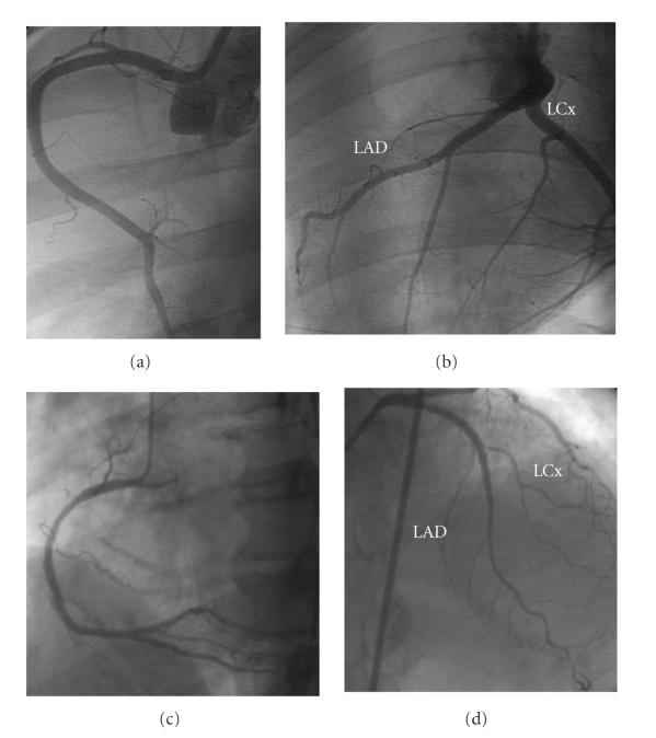
Porcine (a and b). (a) Right coronary artery. (b) Left coronary system. Human (c and d). (c) Right coronary artery. (d) Left coronary system. Similar anatomy and coronary distribution is shown of the left anterior descending, left circumflex, and right coronary arteries.
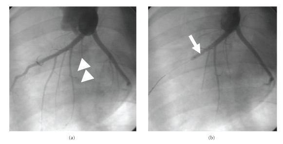
Baseline coronary angiogram was obtained for assessment of anatomic information of the LAD (a). The LAD with the first large diagonal artery (white arrowhead) is delineated. Angiographic confirmation of complete occlusion in the midportions (b) of the LAD with a PTCA balloon catheter (white arrow). Adapted from [12].
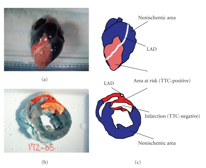
This picture demonstrates the porcine heart (a) and cross-sectional slice (b). The dark area stained with Evans Blue indicates nonischemic territory, and the red area stained with TTC is the area at risk for ischemia (TTC-positive). In (b), the white area is the infarcted (necrotic) myocardium (TTC-negative). The schema explains what the stained colors and territories indicate (c). LAD: left anterior descending artery. Adapted from [12].
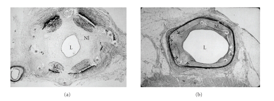
(a) Photomicrographic section shows gross neointimal proliferation causing a significant stenosis (Hematoxylin-Eosin stain, x5). (b) In this section, the porcine coronary was totally occluded with neointimal hyperplasia. L: lumen; M: media; NI: neointima; ∗: stent strut. Adapted from the reference [32].
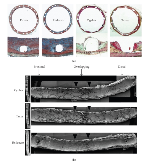
(a) X-rays of longitudinally cut rabbit iliac arteries at 21 days after placement of overlapping ZES, SES, and PES. The extent of stent coverage by endothelial cells was greatest with ZES, with almost complete coverage in the proximal and distal segments and significantly greater coverage in the overlapped segment compared with SES and PES. (b) Photomicrographs showing the amount of neointimal thickness at 28 days after placement of Endeavor zotarolimus-eluting stents (ZESs), Cypher sirolimus-eluting stents (SESs), Taxus paclitaxel-eluting stents (PESs), and Driver bare metal stents (BMSs) in rabbit iliac arteries. With SES, there were focally uncovered stent struts, which were associated with inflammation consisting of heterophils or eosinophils and giant cells. Adapted from [49].
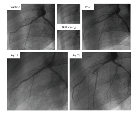
Time course of coronary artery treated with thermal balloon. A severe tandem coronary artery stenosis is observed in the left anterior descending artery (LAD) at 4 weeks after thermal balloon injury [50].
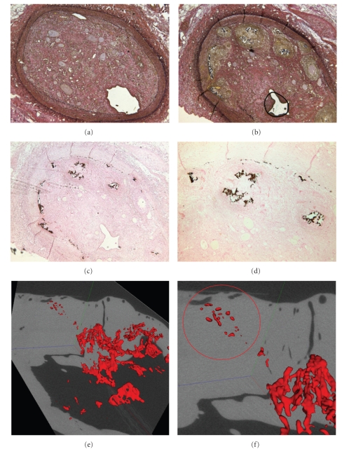
Histological examinations of pig experiments. Elastic-van Gieson (a, b) and Von kossa (c, d) stained CTO segment of coronary arteries. Arrows in (c) and (d) indicate microcalcification. Ex vivo micro-CT confirms that CTO lesions contain calcification in LAD (red circle) [51].
Similar articles
-
A comparative anatomic and physiologic overview of the porcine heart.J Am Assoc Lab Anim Sci. 2014 Sep;53(5):432-8. J Am Assoc Lab Anim Sci. 2014. PMID: 25255064 Free PMC article. Review.
-
Magnetic resonance of the heart and great vessels.Nat Med. 1995 Jul;1(7):711-3. doi: 10.1038/nm0795-711. Nat Med. 1995. PMID: 7585158 Review. No abstract available.
-
Evaluation of the porcine ameroid constrictor model of myocardial ischemia for therapeutic angiogenesis studies.Endothelium. 2006 Jan-Feb;13(1):25-33. doi: 10.1080/10623320600660128. Endothelium. 2006. PMID: 16885064
-
Glycogen Synthase Kinase 3β Inhibition Improves Myocardial Angiogenesis and Perfusion in a Swine Model of Metabolic Syndrome.J Am Heart Assoc. 2016 Jul 12;5(7):e003694. doi: 10.1161/JAHA.116.003694. J Am Heart Assoc. 2016. PMID: 27405812 Free PMC article.
-
Coronary angiography and percutaneous coronary intervention in the porcine model: a practical guide to the procedure.Animal. 2012 Feb;6(2):311-20. doi: 10.1017/S1751731111001650. Animal. 2012. PMID: 22436190
Cited by
-
A fluid-structure interaction model accounting arterial vessels as a key part of the blood-flow engine for the analysis of cardiovascular diseases.Front Bioeng Biotechnol. 2022 Aug 19;10:981187. doi: 10.3389/fbioe.2022.981187. eCollection 2022. Front Bioeng Biotechnol. 2022. PMID: 36061431 Free PMC article.
-
Targeted Delivery for Cardiac Regeneration: Comparison of Intra-coronary Infusion and Intra-myocardial Injection in Porcine Hearts.Front Cardiovasc Med. 2022 Feb 10;9:833335. doi: 10.3389/fcvm.2022.833335. eCollection 2022. Front Cardiovasc Med. 2022. PMID: 35224061 Free PMC article.
-
Role of Coronary Myogenic Response in Pressure-Flow Autoregulation in Swine: A Meta-Analysis With Coronary Flow Modeling.Front Physiol. 2018 May 23;9:580. doi: 10.3389/fphys.2018.00580. eCollection 2018. Front Physiol. 2018. PMID: 29875686 Free PMC article.
-
Pig Induced Pluripotent Stem Cell-Derived Neural Rosettes Parallel Human Differentiation Into Sensory Neural Subtypes.Cell Reprogram. 2017 Apr;19(2):88-94. doi: 10.1089/cell.2016.0057. Epub 2017 Mar 7. Cell Reprogram. 2017. PMID: 28266869 Free PMC article.
-
Domestic Pig (Sus scrofa) as an Animal Model for Experimental Trypanosoma cruzi Infection.Am J Trop Med Hyg. 2016 May 4;94(5):1020-7. doi: 10.4269/ajtmh.15-0233. Epub 2016 Feb 29. Am J Trop Med Hyg. 2016. PMID: 26928841 Free PMC article.
References
-
- Bustad D, Mcclellan D. Swine in biomedical research. Science. 1966;152(3728):1526–1530. - PubMed
-
- Verdouw D P, Van Den Doel MA, De Zeeuw S, Duncker DJ. Animal models in the study of myocardial ischaemia and ischaemic syndromes. Cardiovascular Research. 1998;39(1):121–135. - PubMed
-
- Litvak J, Siderides LE, Vineberg AM. The experimental production of coronary artery insufficiency and occlusion. American Heart Journal. 1957;53(4):505–518. - PubMed
-
- Fozzard HA. Validity of myocardial infarction models. Circulation. 1975;52(6, supplement):131–146. - PubMed
-
- Schwarz ER, Montino H, Fleischhauer J, Klues HG, Vom Dahl J, Hanrath P. The angiotensin II receptor antagonist EXP 3174 reduces infarct size comparable with enalaprilat and augments preconditioning in the pig heart. Cardiovascular Drugs and Therapy. 1997;11(5):687–695. - PubMed
Publication types
MeSH terms
LinkOut - more resources
Full Text Sources