. 2011:2011:920898.
doi: 10.1155/2011/920898. Epub 2010 Dec 22.

Pathobiology of hodgkin lymphoma

Affiliations

Pathobiology of hodgkin lymphoma

Pier Paolo Piccaluga et al. Adv Hematol. 2011.

Abstract

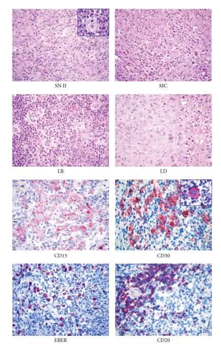
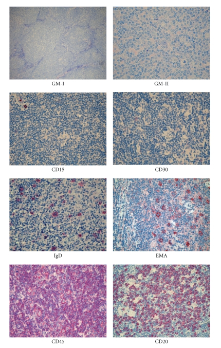
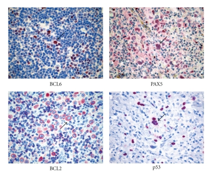
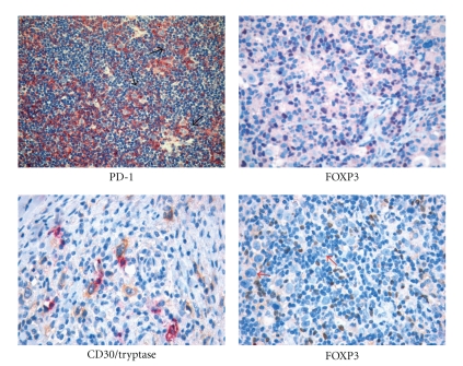
Despite its well-known histological and clinical features, Hodgkin's lymphoma (HL) has recently been the object of intense research activity, leading to a better understanding of its phenotype, molecular characteristics, histogenesis, and possible mechanisms of lymphomagenesis. There is complete consensus on the B-cell derivation of the tumor in most cases, and on the relevance of Epstein-Barr virus infection and defective cytokinesis in at least a proportion of patients. The REAL/WHO classification recognizes a basic distinction between lymphocyte predominance HL (LP-HL) and classic HL (cHL), reflecting the differences in clinical presentation and behavior, morphology, phenotype, and molecular features. cHL has been classified into four subtypes: lymphocyte rich, nodular sclerosing, with mixed cellularity, and lymphocyte depleted. The borders between cHL and anaplastic large-cell lymphoma have become sharper, whereas those between LP-HL and T-cell-rich B-cell lymphoma remain ill defined. Treatments adjusted to the pathobiological characteristics of the tumor in at-risk patients have been proposed and are on the way to being applied.


Figures

Figure 1
Figure 1
Histopathological features of classical Hodgkin lymphoma (cHL). At morphology (H&E staining), it is possible to distinguish nodular sclerosis (NS)*, mixed cellularity (MC), lymphocyte rich (LR), and lymphocyte-depleted (LD) subtypes. At immunophenotyping, cHL is typically CD15+, CD30+∗, possibly EBER+, and CD20-/+ (Olympus BX41 microscope, Olympus CAMEDIA C-7070 camera, magnification ×400, colours balanced after acquisition with Adobe Photoshop). *Note the typical Reed-Sternberg cells in the insets.
Figure 2
Figure 2
Histopathological features of lymphocyte predominant Hodgkin lymphoma (LP-HL). At morphology (GIEMSA staining), it is possible to appreciate the nodular morphology (GM-I) and the typical LP cells (GM-II, arrow). At immunophenotyping, it is typically CD15, CD30, EMA+, CD45+, CD20+, and possibly (10%–20% cases) IgD+ (Olympus BX41 microscope, Olympus CAMEDIA C-7070 camera, magnification ×400, colours balanced after acquisition with Adobe Photoshop).
Figure 3
Figure 3
Immunophenotyping of Hodgkin lymphoma. Immunostains for BCL6, PAX5, BCL2, and p53 are shown. Please note positive staining in the diagnostic cells (arrows).
Figure 4
Figure 4
The reactive “milieau” in Hodgkin lymphoma. Mast cells and regulatory T cells populate the HL microenvironment showing spatial interaction with RS cells. Immunohistochemical staining for PD-1 and FOXP3 highlights the presence of several regulatory T cells intermingling with RS cells (arrows). Double immunohistochemistry for CD30 (yellow/brown) and mast cell tryptase (purple) shows the tight interaction of mast cells with RS cells.
Figure 5
Figure 5
Schematic representation of the reciprocal contribution of mast cells (MC), effector T cells (Teff), and regulatory T cells (Treg) to the HL-associated immunological microenvironment. (a) Mast cells, as other innate immune players directly sustain RS cell proliferation and proinflammatory mediator synthesis. (b) This effect is boosted by Teff that contributes to mast cell activation and amplifies the inflammatory spur. (c) Treg can interfere with the activation status of both Teff and MC through the OX40/OX40L axis and TGF-β release eventually limiting the delivery of the proliferation/survival signal to RS cells. (d) However, when the microenvironment is diverted towards marked inflammation owing to the abundant presence of activated MC, the regulatory function of Treg may prove inadequate to restore the balance between pro- and anti-inflammatory stimuli, and Treg can even boost inflammation through TGF-β release and Th17 generation.

References

    1. Stein H, Delsol G, Pileri S, Weiss L, Poppema S, Jaffe E. Classical Hodgkin lymphoma, introduction. In: Swerdlow S, Campo E, Harris NL, et al., editors. WHO Classification of Tumors of the Hematopoietic and Lymphoid Tissue. Lyon, France: IARC; 2008. pp. 326–329.
    1. Hummel M, Marafioti T, Ziemann K, Stein H. Ig rearrangements in isolated Reed-Sternberg cells: conclusions from four different studies. Annals of Oncology. 1996;7(supplement 4):S31–S33. - PubMed
    1. Marafioti T, Hummel M, Anagnostopoulos I, et al. Origin of nodular lymphocyte-predominant Hodgkin’s disease from a clonal expansion of highly mutated germinal-center B cells. New England Journal of Medicine. 1997;337(7):453–458. - PubMed
    1. Izban KF, Nawrocki JF, Alkan S, Hsi ED. Monoclonal IgH gene rearrangement in microdissected nodules from nodular sclerosis Hodgkin disease. American Journal of Clinical Pathology. 1998;110(5):599–606. - PubMed
    1. Bräuninger A, Hansmann ML, Strickler JG, et al. Identification of common germinal-center B-cell precursors in two patients with both Hodgkin’s disease and non-Hodgkin’s lymphoma. New England Journal of Medicine. 1999;340(16):1239–1247. - PubMed