. 2011 Jan 6;6(1):e15976.
doi: 10.1371/journal.pone.0015976.

Deciphering human heat shock transcription factor 1 regulation via post-translational modification in yeast

Affiliations

Deciphering human heat shock transcription factor 1 regulation via post-translational modification in yeast

Liliana Batista-Nascimento et al. PLoS One. .

Abstract

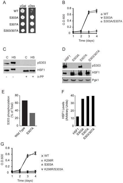
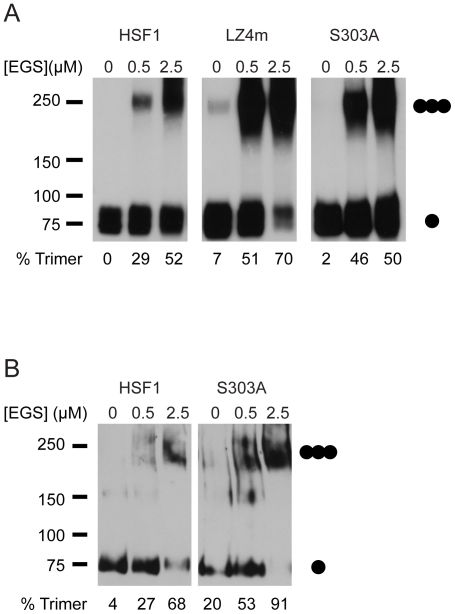
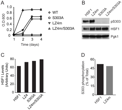
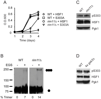
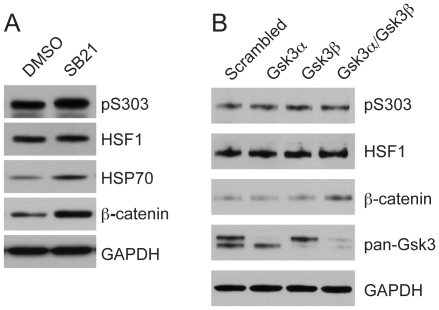
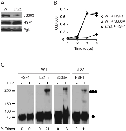
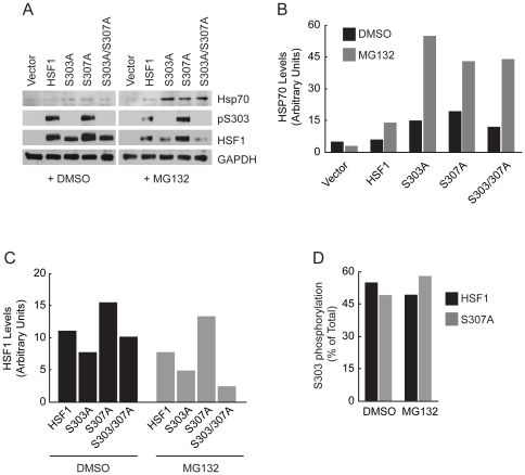
Heat shock transcription factor 1 (HSF1) plays an important role in the cellular response to proteotoxic stresses. Under normal growth conditions HSF1 is repressed as an inactive monomer in part through post-translation modifications that include protein acetylation, sumoylation and phosphorylation. Upon exposure to stress HSF1 homotrimerizes, accumulates in nucleus, binds DNA, becomes hyper-phosphorylated and activates the expression of stress response genes. While HSF1 and the mechanisms that regulate its activity have been studied for over two decades, our understanding of HSF1 regulation remains incomplete. As previous studies have shown that HSF1 and the heat shock response promoter element (HSE) are generally structurally conserved from yeast to metazoans, we have made use of the genetically tractable budding yeast as a facile assay system to further understand the mechanisms that regulate human HSF1 through phosphorylation of serine 303. We show that when human HSF1 is expressed in yeast its phosphorylation at S303 is promoted by the MAP-kinase Slt2 independent of a priming event at S307 previously believed to be a prerequisite. Furthermore, we show that phosphorylation at S303 in yeast and mammalian cells occurs independent of GSK3, the kinase primarily thought to be responsible for S303 phosphorylation. Lastly, while previous studies have suggested that S303 phosphorylation represses HSF1-dependent transactivation, we now show that S303 phosphorylation also represses HSF1 multimerization in both yeast and mammalian cells. Taken together, these studies suggest that yeast cells will be a powerful experimental tool for deciphering aspects of human HSF1 regulation by post-translational modifications.


Conflict of interest statement

Competing Interests: The authors have declared that no competing interests exist.

Figures

Figure 1
Figure 1. S303 phosphorylation represses HSF1 activity in yeast.
(A) PS145 yeast strains expressing wild-type HSF1 (WT) or the S303A, S307A or S303/307A mutants were plated on either galactose or dextrose supplemented medium. (B) PS145 expressing either wild-type HSF1 or the S303A or S303/307A mutants were grown in dextrose containing medium for 4 d. Growth was monitored by measuring O.D.600. (C) HeLa cells were grown at 37°C (C) or heat shocked for 2 h at 42°C (HS). Total protein extracts were treated with lambda protein phosphatase and analyzed for phospho-S303 (pS303) and total HSF1 levels by immunoblotting. (D) PS145 was transformed with a plasmid expressing wild-type HSF1 (WT) or mutant alleles of HSF1 and grown on galactose containing medium. Total protein extracts were analyzed for pS303, HSF1 and Pgk1 by immunoblotting. (E) Levels of HSF1 phosphorylated at S303 were quantified and are shown as a percent of total HSF1, from panel D. (F) Protein levels of HSF1 were normalized to Pgk1, from panel D. (G) PS145 expressing either wild-type HSF1 or mutant HSF1 alleles were assayed for HSF1-dependent growth as in B.
Figure 2
Figure 2. S303 represses trimer formation of HSF1 in yeast and mammalian cells.
(A) PS145 was transformed with wild-type HSF1, the LZ4m mutant or the S303A mutant and grown on galactose containing medium. Total protein extracts were evaluated for HSF1 multimerization by EGS crosslinking, SDS-PAGE, and immunoblotting using an HSF1 specific antibody. The positions of molecular weight markers are indicated on the left, and circles indicating the expected migration of HSF1 monomers and trimers are on the right. Levels of HSF1 trimer as percent of total HSF1 are shown below. (B) hsf1−/− MEFs were transfected with a plasmid expressing wild-type HSF1 or the S303A mutant and analyzed for HSF1 multimerization by EGS cross-linking as in A.
Figure 3
Figure 3. Phosphorylation of S303 and coiled-coil domains synergize in the repression of HSF1 in yeast.
(A) PS145 expressing either wild-type HSF1 or mutant alleles of HSF1 were grown in dextrose supplemented medium for 4 d. Growth was monitored by measuring O.D.600. (B) PS145 was transformed with a plasmid expressing wild-type HSF1 (WT) or mutant alleles of HSF1 and grown on galactose containing medium. Total protein extracts were analyzed for pS303, total HSF1 and Pgk1 by immunoblotting. (C) Protein levels of HSF1 were normalized to Pgk1, from panel B. (D) Levels of HSF1 phosphorylated at S303 were quantified and are shown as a percent of total HSF1, from panel B.
Figure 4
Figure 4. GSK3 represses HSF1 activity in yeast independent of S303.
(A) PS145 (WT) expressing wild-type HSF1 or the S303A HSF1 mutant and LNY1 (rim11Δ) expressing wild-type HSF1 were grown in dextrose supplemented medium for 4 d. Growth was monitored by measuring O.D.600. (B) PS145 (WT) and LNY1 (rim11Δ) expressing wild-type HSF1 were grown on galactose containing medium and were evaluated for HSF1 multimerization by EGS crosslinking, SDS-PAGE, and immunoblotting using an HSF1 specific antibody. The positions of molecular weight markers are indicated on the left, and circles indicating the expected migration of HSF1 monomers and trimers are on the right. Levels of HSF1 trimer as percent of total HSF1 are shown below. (C) PS145 (WT) and LNY1 (rim11Δ) were transformed with a plasmid expressing wild-type HSF1 and were grown on galactose containing medium. Total protein extracts were analyzed for pS303, total HSF1 and Pgk1 by immunoblotting. (D) YPH499 (WT) and LNY3 (4xgsk3Δ) were transformed with a plasmid expressing wild-type HSF1 and were grown in dextrose containing medium. Total protein extracts were analyzed for pS303, total HSF1 and Pgk1 by immunoblotting.
Figure 5
Figure 5. GSK3 represses HSF1 activity in HeLa cells independent of S303 phosphorylation.
(A) HeLa cells were treated with DMSO solvent or the GSK3 inhibitor SB-216763 (25 µM) for 15 h. Total protein was analyzed for pS303, HSF1, and β-catenin by immunoblotting. GAPDH serves as a loading control. (B) HeLa cells were treated with siRNA specific for GSK3α and GSK3β either individually or together or a scrambled siRNA for 72 h. Total protein was analyzed for pS303, total HSF1, β-catenin, GSK3α/β and GAPDH by immunoblotting.
Figure 6
Figure 6. S303 phosphorylation of HSF1 in yeast is modulated by Slt2.
(A) PS145 and LNY2 (slt2Δ) were transformed with a plasmid expressing wild-type HSF1 and were grown on galactose containing medium. Total protein extracts were analyzed for pS303, total HSF1 and Pgk1 by immunoblotting. (B) PS145 (WT) expressing wild-type HSF1 or the S303A mutant or LNY2 (slt2Δ) expressing wild-type HSF1 were grown in dextrose supplemented medium for 4 d. Growth was monitored by measuring O.D.600. (C) PS145 (WT) expressing wild-type HSF1, the LZ4m mutant or the S303A mutant and LNY2 (slt2Δ) expressing HSF1 were evaluated for HSF1 multimerization by EGS cross-linking, SDS-PAGE, and immunoblotting. The positions of molecular weight markers are indicated on the left and circles indicating the expected migration of HSF1 monomers and trimers are on the right. Levels of HSF1 trimer as percent of total HSF1 are shown below.
Figure 7
Figure 7. S303 and S307 repress HSF1 activity in hsf1−/− MEFs.
(A) hsf1−/− MEFs were transfected with an empty vector or plasmids expressing wild-type HSF1 or the S303A, S307A or the S303/307A mutants. The transfected cells were treated with DMSO solvent or MG132 (10 µM) for 5 h. Total protein extracts were analyzed for Hsp70, pS303 and HSF1 by immunoblotting. GAPDH serves as a loading control. (B) Protein levels of Hsp70 were normalized to GAPDH, from panel A. (C) Protein levels of HSF1 were normalized to GAPDH, from panel A. (D) Levels of HSF1 phosphorylated at S303 were quantified and are shown as a percent of total HSF1, from panel A.

References

    1. Morimoto RI. Proteotoxic stress and inducible chaperone networks in neurodegenerative disease and aging. Genes Dev. 2008;22:1427–1438. - PMC - PubMed
    1. Akerfelt M, Morimoto RI, Sistonen L. Heat shock factors: integrators of cell stress, development and lifespan. 2010. Nature reviews Molecular cell biology. - PMC - PubMed
    1. Hahn J-S, Hu Z, Thiele DJ, Iyer VR. Genome-wide analysis of the biology of stress responses through heat shock transcription factor. Mol Cell Biol. 2004;24:5249–5256. - PMC - PubMed
    1. Farkas T, Kutskova YA, Zimarino V. Intramolecular repression of mouse heat shock factor 1. Mol Cell Biol. 1998;18:906–918. - PMC - PubMed
    1. Orosz A, Wisniewski J, Wu C. Regulation of Drosophila heat shock factor trimerization: global sequence requirements and independence of nuclear localization. Mol Cell Biol. 1996;16:7018–7030. - PMC - PubMed

Publication types

MeSH terms