. 2011 Jan 6;6(1):e15967.
doi: 10.1371/journal.pone.0015967.

Elimination of hepatitis C virus from hepatocytes by a selective activation of therapeutic molecules

Affiliations

Elimination of hepatitis C virus from hepatocytes by a selective activation of therapeutic molecules

Xiaoyu Wen et al. PLoS One. .

Abstract

To eliminate hepatitis C virus (HCV) from infected hepatocytes, we generated two therapeutic molecules specifically activated in cells infected with HCV. A dominant active mutant of interferon (IFN) regulatory factor 7 (IRF7) and a negative regulator of HCV replication, VAP-C (Vesicle-associated membrane protein-associated protein subtype C), were fused with the C-terminal region of IPS-1 (IFNβ promoter stimulator-1), which includes an HCV protease cleavage site that was modified to be localized on the ER membrane, and designated cIRF7 and cVAP-C, respectively. In cells expressing the HCV protease, cIRF7 was cleaved and the processed fragment was migrated into the nucleus, where it activated various IFN promoters, including promoters of IFNα6, IFNβ, and IFN stimulated response element. Activation of the IFN promoters and suppression of viral RNA replication were observed in the HCV replicon cells and in cells infected with the JFH1 strain of HCV (HCVcc) by expression of cIRF7. Suppression of viral RNA replication was observed even in the IFN-resistant replicon cells by the expression of cIRF7. Expression of the cVAP-C also resulted in suppression of HCV replication in both the replicon and HCVcc infected cells. These results suggest that delivery of the therapeutic molecules into the liver of hepatitis C patients, followed by selective activation of the molecules in HCV-infected hepatocytes, is a feasible method for eliminating HCV.


Conflict of interest statement

Competing Interests: The authors have declared that no competing interests exist.

Figures

Figure 1
Figure 1. Dominant active mutant of IRF7 activates IFN promoters in cells replicating HCV.
(A) Structures of IRF3, IRF7 and the dominant active mutants, IRF3m and IRF7m. The DNA-binding domain, nuclear export sequence, transactivation domain, association domain, inhibitory domain, and signal response domain are indicated as DBD, NES, TAD, AD, ID, and SRD, respectively. Huh7 cells and HCV replicon cells (1×10⁵ cells/well) (B), and Huh7OK1 cells (7.5×10⁴ cells/well) infected with HCVcc at an moi of 1 and incubated for 72 h (C) were transfected with 100 ng of plasmid encoding the luciferase gene under the control of the IFNα6, IFNβ, or ISRE promoter together with an empty vector (EV) or a plasmid encoding each of the IRF constructs. The relative luciferase activity of cell lysates was determined at 24 h post-transfection. HCV replicon cells (3×10⁵ cells/well) (D) and Huh7OK1 cells (1.5×10⁵ cells/well) infected with HCVcc at an moi of 1 and incubated for 72 h (E) were transfected with EV or a plasmid encoding each of the IRF constructs and the expressions of NS5A, IRFs, and GAPDH (upper panel) and synthesis of viral RNA (lower panel) at 72 h post-transfection were determined by immunoblotting and real-time PCR after standardization with GAPDH, respectively. The data shown in this figure are representative of three independent experiments. The error bars represent the standard deviations. Asterisks indicate significant differences (**P<0.01) versus the control cells or mock-infected cells.
Figure 2
Figure 2. Construction of cIRF7 capable of activating the IFN promoters in cells replicating HCV.
(A top) Schematic representation of the cIRF7 constructs. cIRF7 consists of IRF7m, FLAG-tag, and IPS-1 (503 to 540 amino acid residues) sequences containing a cleavage site by HCV NS3/4A protease, a transmembrane domain and a cytoplasmic region modified to localize on the ER. cIRF7(C508A) has a substitution of Cys508 to Ala which renders it resistant to the cleavage by the HCV protease. (A bottom) Immunoblot analyses of 293T cells transfected with a plasmid encoding either cIRF7 or cIRF7(C508A) together with either an empty vector (EV) or a plasmid encoding either FLAG-tagged HCVNS3/4A or FLAG-tagged HCVNS3/4A (S139A). (B) 293T cells (2×10⁵ cells/well) were transfected with a plasmid of EV, FLAG-tagged HCVNS3/4A or FLAG-tagged HCVNS3/4A(S139A) in combination with a plasmid of EV, cIRF7 or cIRF7 (C508A) together with 100 ng of the reporter plasmid encoding the luciferase gene under the control of the IFNα6, IFNβ or ISRE promoter, and luciferase activity was determined at 24 h post-transfection. (C) HCV replicon cells (1.5×10⁵ cells/well) and (D) Huh7OK1 cells (7.5×10⁴ cells/well) infected with HCVcc at an moi of 1 and incubated for 72 h were transfected with 100 ng of each of the reporter plasmids together with plasmid of EV, cIRF7 or cIRF7(C508A) and luciferase activity was determined at 24 h post-transfection. (E) Huh7 cells, HCV subgenomic replicon cells, and JEV subgenomic replicon cells (1×10⁵ cells/well) (top) and Huh7OK1 cells (7.5×10⁴ cells/well) infected with JEV and HCV (bottom) at an moi of 0.008, 0.04, 0.2, and 1 and incubated for 24 h and 72 h, respectively, were transfected with 100 ng of each of the reporter plasmids together with cIRF7 and the luciferase activity was determined at 24 h post-transfection. The data shown in this figure are representative of three independent experiments. The error bars represent the standard deviations. Asterisks indicate significant differences (*P<0.05, **P<0.01) versus the control cells or mock-infected cells.
Figure 3
Figure 3. Scheme of activation of the therapeutic molecule in cells infected with HCV.
The chimeric molecules are cleaved by HCV NS3/4A protease and the released fragments inhibit propagation of HCV through induction of IFN after translocation into the nucleus (cIRF7) or disruption of the replication complex (cVAP-C), whereas the molecule is stably anchored in the ER within uninfected cells.
Figure 4
Figure 4. Specificity of activation of the IFN promoters by the expression of cIRF7.
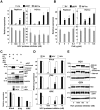
(A) HCV replicon cells (1.5×10⁵ cells/well) or Huh7OK1 cells (7.5×10⁴ cells/well) infected with HCVcc at an moi of 1 and incubated for 72 h were treated with various concentrations of HCV protease inhibitor (A) or cyclosporine A (CsA) (B), transfected with an empty vector (EV) (white bars) or plasmids encoding cIRF7 (black bars) or IRF7m (gray bars) together with 100 ng of a reporter plasmid encoding the luciferase gene under the control of the ISRE promoter, and luciferase activity was determined at 24 h post-transfection. (C top) A plasmid encoding cIRF7 was co-transfected with a plasmid encoding either FLAG-tagged HCVNS3/4A, FLAG-tagged GBVNS3/4A, or HA-tagged JEVNS2b/3 into 293T cells, and the expressions of cIRF7, viral proteases and GAPDH were determined by immunoblotting. (C bottom) 293T cells (2×10⁵ cells/well) transfected with a plasmid encoding either EV (dark gray bars), FLAG-tagged HCVNS3/4A (black bars), FLAG-tagged GBVNS3/4A (white bars), or HA-tagged JEVNS2b/3 (gray bars) together with 100 ng of the plasmid encoding the luciferase gene under the control of the promoter of either IFNα6, IFNβ or ISRE, and luciferase activity was determined at 24 h post-transfection. (D) 293T cells (2×10⁵ cells/well) were transfected with 100 ng of the reporter plasmids together with plasmids encoding EV (gray bars), FLAG-tagged HCVNS3/4A (black bars) or FLAG-tagged GBVNS3/4A (white bars) in the presence or absence of the HCV protease inhibitor, and luciferase activity was determined at 24 h post-transfection. (E) cIRF7 was co-expressed with FLAG-tagged HCVNS3/4A or FLAG-tagged GBVNS3/4A in 293T cells in the presence or absence of the HCV protease inhibitor, and the expressions of cIRF7, viral proteases and GAPDH were determined by immunoblotting. The data shown in this figure are representative of three independent experiments. The error bars represent the standard deviations. Asterisks indicate significant differences (*P<0.05, **P<0.01) versus the control cells or mock-infected cells.
Figure 5
Figure 5. Activation of cIRF7 in cells expressing HCV protease.
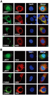
(A) Huh7OK1 cells (5×10⁴ cells/well) were co-transfected with plasmids encoding either EGFP-cIRF7 or EGFP-cIRF7(C508A) and plasmids encoding either HCVNS3/4A, HCVNS3/4A(S139A) or NS5A, harvested at 24 h post-transfection, fixed with 4% paraformaldehyde in PBS, and permeabilized with 0.25% saponin. HCV NS3 and NS5A were stained with the appropriate antibodies, followed by staining with AF594-conjugated second antibodies. (B) HCV replicon cells (5×10⁴ cells/well) were transfected with plasmids encoding either EGFP-cIRF7 or EGFP-cIRF7(C508A), and endogenous expression of HCV NS3 and an ER marker, PDI, was detected in cells treated and stained with the appropriate antibodies as described above. Subcellular localization of cIRF7s, HCV proteins and PDI was determined by confocal microscopy after staining of nuclei by DAPI. The data shown in this figure are representative of three independent experiments.
Figure 6
Figure 6. Suppression of HCV replication by the expression of cIRF7.
(A) HCV replicon cells (3×10⁵ cells/well) and (B) Huh7OK1 cells (1.5×10⁵ cells/well) infected with HCVcc at an moi of 1 and incubated for 72 h were transfected with a plasmid encoding either empty vector (EV), cIRF7 or cIRF7(C508A), and the expression of NS5A, cIRF7s and GAPDH (upper panels) and synthesis of viral RNA (lower panels) were determined at 72 h post-transfection by immunoblotting and real-time PCR, respectively. (C upper left) HCV Con1 replicon cells and 4βR replicon cells exhibiting an IFN-resistant phenotype (1.5×10⁵ cells/well) were treated with the CsA (5 µg/ml) or 104, 103, and 102 units/ml of recombinant human IFNα and the expressions of NS5A and GAPDH were determined by immunoblotting. The 4βR replicon cells (3×10⁵ cells/well) were transfected with EV or plasmid encoding either cIRF7 or cIRF7(C508A), and the expressions of NS5A, cIRF7s and GAPDH (C upper right) and synthesis of viral RNA (C lower right) were determined at 72 h post-transfection by immunoblotting and real-time PCR, respectively. The 4βR cells and their cured cells (4βRc) with the HCV genome eliminated (1×10⁵ cells/well) were transfected with EV or plasmid encoding either cIRF7 or cIRF7(C508A) together with 100 ng of plasmid encoding the luciferase gene under the control of the ISRE promoter, and luciferase activity was determined at 24 h post-transfection (C lower left). The data shown in this figure are representative of three independent experiments. The error bars represent the standard deviations. Asterisks indicate significant differences (*P<0.05, **P<0.01) versus the control cells or mock-infected cells.
Figure 7
Figure 7. Suppression of HCV replication by the expression of cVAP-C.
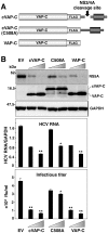
(A) Schematic representation of cVAP-C, cVAP-C(C508A) and VAP-C. Like cIRF7, cVAP-C is composed of the sequences of VAP-C, FLAG-tag, and the C-terminus domain of IPS-1. (B) Huh7OK1 cells (1.5×10⁵ cells/well) infected with HCVcc at an moi of 1 and incubated for 72 h were transfected with EV, or plasmid encoding either cIRF7 or cIRF7(C508A), and the expressions of NS5A, VAP-Cs and GAPDH (top panel), synthesis of viral RNA (middle panel) and infectious titers in the culture supernatants were determined at 72 h post-transfection by immunoblotting, real-time PCR, and focus forming assay, respectively. The data shown in this figure are representative of three independent experiments. The error bars represent the standard deviations. Asterisks indicate significant differences (*P<0.05, **P<0.01) versus the control cells or mock-infected cells.

Similar articles

Cited by

References

    1. Cerny A, Chisari FV. Pathogenesis of chronic hepatitis C: immunological features of hepatic injury and viral persistence. Hepatology. 1999;30:595–601. - PubMed
    1. Moriishi K, Matsuura Y. Mechanisms of hepatitis C virus infection. Antivir Chem Chemother. 2003;14:285–297. - PubMed
    1. Fried MW. Side effects of therapy of hepatitis C and their management. Hepatology. 2002;36:S237–244. - PubMed
    1. Wakita T, Pietschmann T, Kato T, Date T, Miyamoto M, et al. Production of infectious hepatitis C virus in tissue culture from a cloned viral genome. Nat Med. 2005;11:791–796. - PMC - PubMed
    1. Sen GC. Viruses and interferons. Annu Rev Microbiol. 2001;55:255–281. - PubMed

Publication types

MeSH terms

Substances