

Targeting skin dendritic cells to improve intradermal vaccination
- PMID: 21253784
- PMCID: PMC4285659
- DOI: 10.1007/82_2010_118
Targeting skin dendritic cells to improve intradermal vaccination
Abstract
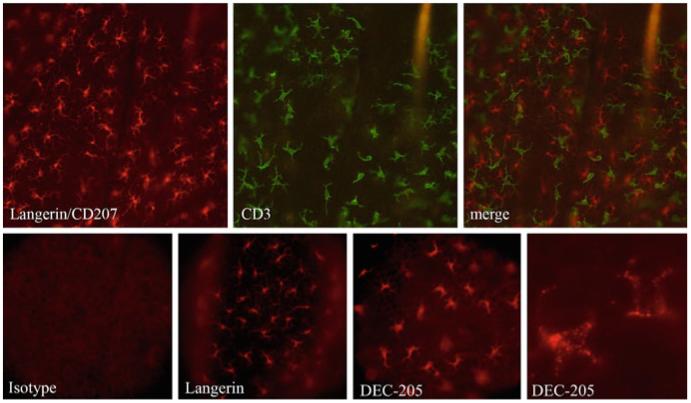
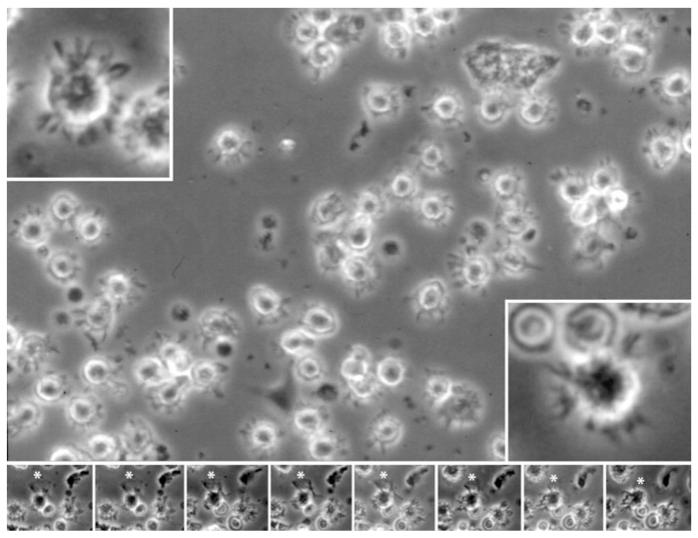
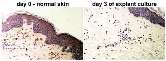
Vaccinations in medicine are typically administered into the muscle beneath the skin or into the subcutaneous fat. As a consequence, the vaccine is immunologically processed by antigen-presenting cells of the skin or the muscle. Recent evidence suggests that the clinically seldom used intradermal route is effective and possibly even superior to the conventional subcutaneous or intramuscular route. Several types of professional antigen-presenting cells inhabit the healthy skin. Epidermal Langerhans cells (CD207/langerin(+)), dermal langerin(neg), and dermal langerin(+) dendritic cells (DC) have been described, the latter subset so far only in mouse skin. In human skin langerin(neg) dermal DC can be further classified based on their reciprocal expression of CD1a and CD14. The relative contributions of these subsets to the generation of immunity or tolerance are still unclear. Yet, specializations of these different populations have become apparent. Langerhans cells in human skin appear to be specialized for induction of cytotoxic T lymphocytes; human CD14(+) dermal DC can promote antibody production by B cells. It is currently attempted to rationally devise and improve vaccines by harnessing such specific properties of skin DC. This could be achieved by specifically targeting functionally diverse skin DC subsets. We discuss here advances in our knowledge on the immunological properties of skin DC and strategies to significantly improve the outcome of vaccinations by applying this knowledge.
Figures
References
-
- Aberer W, Kruisbeek AM, Shimada S, Katz SI. In vivo treatment with anti-I-A antibodies: differential effects on Ia antigens and antigen-presenting cell function of spleen cells and epidermal Langerhans cells. J Immunol. 1986;136:830–836. - PubMed
-
- Allam JP, Niederhagen B, Bucheler M, Appel T, Betten H, Bieber T, Berge S, Novak N. Comparative analysis of nasal and oral mucosa dendritic cells. Allergy. 2006;61:166–172. - PubMed
-
- Allan RS, Smith CM, Belz GT, Van Lint AL, Wakim LM, Heath WR, Carbone FR. Epidermal viral immunity induced by CD8α+ dendritic cells but not by Langerhans cells. Science. 2003;301:1925–1928. - PubMed
-
- Angel CE, Lala A, Chen CJ, Edgar SG, Ostrovsky LL, Dunbar PR. CD14+ antigen-presenting cells in human dermis are less mature than their CD1a+ counterparts. Int Immunol. 2007;19:1271–1279. - PubMed
Publication types
MeSH terms
Substances
Grants and funding
LinkOut - more resources
Full Text Sources
Other Literature Sources
Medical
Research Materials
