. 2011 Jan;53(1):73-85.
doi: 10.1002/hep.23977. Epub 2010 Oct 26.

Hypercytolytic activity of hepatic natural killer cells correlates with liver injury in chronic hepatitis B patients

Affiliations

Hypercytolytic activity of hepatic natural killer cells correlates with liver injury in chronic hepatitis B patients

Zheng Zhang et al. Hepatology. 2011 Jan.

Abstract

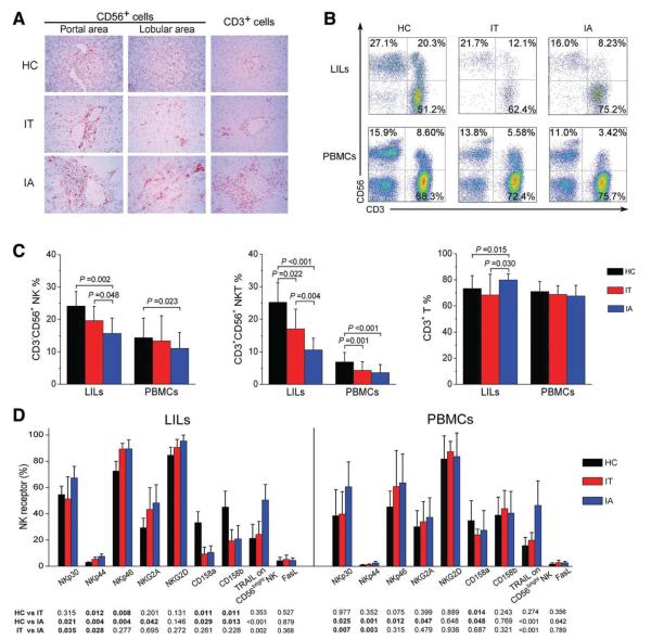
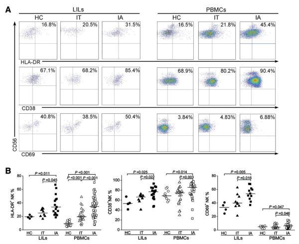
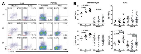
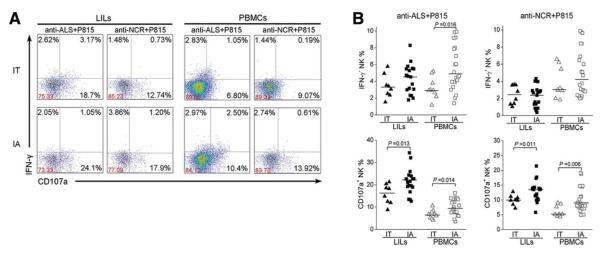
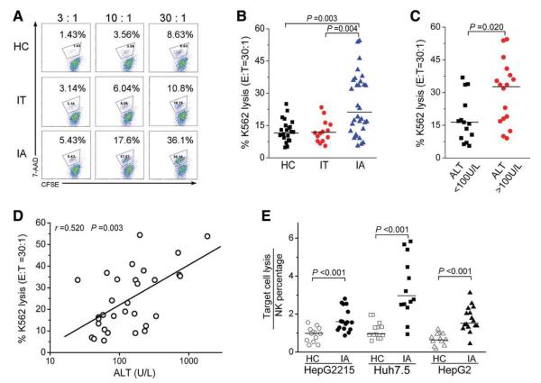
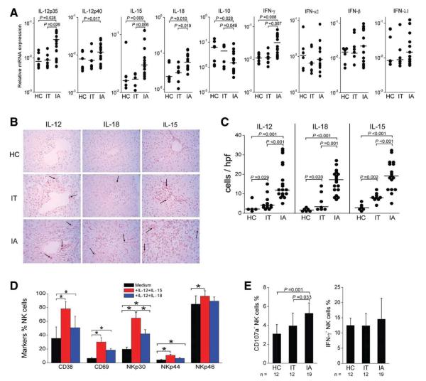
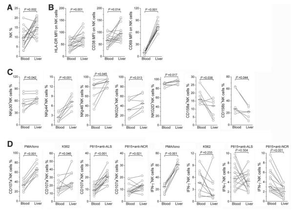
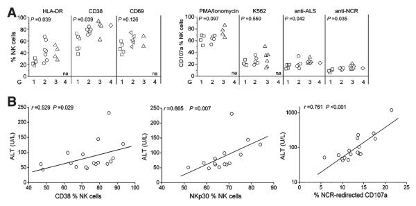
Natural killer (NK) cells are abundant in the liver and serve as a major innate immune component against microbial infection. Although NK cells have been implicated in inducing hepatocellular damage in patients with chronic hepatitis virus infections, the roles that hepatic NK cells play in chronic hepatitis B virus (HBV) infections remain obscure. In this study, we comprehensively characterized intrahepatic and peripheral NK cells and investigated their impact on liver pathology in a cohort of HBV-infected individuals; this cohort included 51 immune-activated (IA) patients, 27 immune-tolerant (IT) carriers, and 26 healthy subjects. We found that NK cells expressing NK receptors (activation receptors) preferentially accumulated in the livers of IA patients, in which they were activated and skewed toward cytolytic activity but without a concomitant increase in interferon-γ production, in comparison with those of IT carriers and healthy subjects. Further analysis showed that the livers of IA patients, in comparison with those of IT and healthy subjects, expressed higher levels of interleukin-12 (IL-12), IL-15, and IL-18 in situ and lower levels of IL-10, which in vitro can induce the activation and degranulation of NK cells from healthy individuals. Finally, hepatic NK cells displayed more cytolytic activity than peripheral NK cells, and this was found to be positively correlated with the liver histological activity index and serum alanine aminotransferase levels in these IA patients.

Conclusion: In IA patients, hepatic NK cells are activated and preferentially skew toward cytolytic activity, which depends on an imbalanced cytokine milieu and correlates with liver injury during chronic HBV infection.


Figures

Fig. 1
Fig. 1
Activation receptor–expressing NK cells selectively accumulate in the livers of IA patients with chronic HBV infection. (A) Immunohistochemical detection of CD56+ and CD3+ cells in the liver tissues of HBV-infected IA patients, IT subjects, and HC donors (magnification, 400 ×). Positively stained cells appear red and were present in both portal and lobular areas in the livers. (B) Representative dot plots of CD3+ and CD56+ staining in LILs and PBMCs isolated from HC subjects and HBV-infected individuals are shown. Values in the quadrants represent the percentages of CD3CD56+ NK cells, CD3+CD56+ NKT cells, and CD3+ T cells. (C) Summarized data show the percentages of NK cells, NKT cells, and T cells in the IA (n = 51), IT (n = 27), and HC groups (n = 26). (D) Pooled data show the percentages of hepatic and peripheral NK cells expressing NK receptors, TRAIL, and FasL from the three indicated groups. The data are shown as means and standard deviations. P values are shown.
Fig. 2
Fig. 2
NK cells express high levels of activation markers in IA patients. (A) Representative dot plots depict the expression of the activation markers HLA-DR, CD38, and CD69 on both hepatic and peripheral NK cells from IA patients and IT and HC subjects. CD3CD56+ NK cells were gated. Values in the upper right quadrants represent the percentages of CD3CD56+ NK cells expressing activation markers. (B) Pooled data show the percentages of hepatic and peripheral CD3CD56+ NK cells expressing HLA-DR (left), CD38 (middle), and CD69 (right) among the three groups of subjects.
Fig. 3
Fig. 3
NK cells from IA patients are prone to degranulation. (A) Representative dot plots depict IFN-γ and CD107a expression on both hepatic and peripheral NK cells among the three groups in response to various stimuli. CD3CD56+ NK cells were gated. Values in the quadrants represent the percentages of CD3CD56+ NK cells expressing IFN-γ and CD107a. (B) Pooled data show the percentages of hepatic and peripheral CD3CD56+ NK cells expressing IFN-γ and CD107a among the three groups upon stimulation with PMA/ionomycin and K562 cells. Each dot represents one individual.
Fig. 4
Fig. 4
Redirected cytotoxicity of NK cells is increased in IA patients. (A) Representative dot plots depict the redirected NK degranulation assay using P815 target cells and anti-ALS (anti-CD16) and anti-NCR antibodies in LILs and PBMCs from IA patients and IT subjects. CD3CD56+ NK cells were gated. Values in the quadrants represent the percentages of CD3CD56+ NK cells expressing IFN-γ and CD107a. (B) Pooled data show the percentages of hepatic and peripheral CD3CD56+ NK cells expressing IFN-γ and CD107a from IA patients and IT subjects in response to anti-ALS and anti-NCR antibodies. Each dot represents one individual.
Fig. 5
Fig. 5
NK cells from IA patients exhibit increased cytolytic activity. (A) Representative dot plots show K562 lysis by PBMCs from the three groups at different E:T ratios (3:1, 10:1, and 30:1). Values in the quadrant represent the percentages of K562 lysis. (B) Pooled data show the percentages of K562 lysis by PBMCs from the three groups at the E:T ratio of 30:1. (C) Pooled data show the percentages of K562 lysis by PBMCs from IA patients with high levels (>100 U/L) or low levels (<100 U/L) of serum ALT. (D) Correlation analysis of the percentages of K562 lysis by PBMCs at the E:T ratio of 30:1 and the serum ALT levels in IA patients (n = 31). (E) Pooled data show the lysis efficiency of hepatocellular carcinoma cell lines (including Huh7.5, HepG2, and HepG2215 cells) by PBMCs from IA patients at the E:T ratio of 30:1. The y axis indicates the ratio of target lysis by NK percentages.
Fig. 6
Fig. 6
Evidence for the contribution of IL-12, IL-15, and IL-18 to increased NK cytolytic activity in IA patients. (A) Statistical analysis of the mRNA expression of hepatic cytokines in IA patients (n = 15) and in IT (n = 6) and HC subjects (n = 8). (B) Immunohistochemical detection of IL-12p70+, IL-15+, and IL-18+ cells in the liver tissues of HBV-infected IA= patients, IT subjects, and HC donors (magnification, 400 ×). Positively stained cells appear red and were present in the livers. (C) Quantitative analysis of cytokine-positive cells in the livers of these subjects. Each dot represents one individual. The horizontal bars indicate the median percentiles. The significant P values are shown. (D) IL-12 in combination with IL-15 or IL-18 induced NK cell activation in vitro, as indicated by CD38 and CD69 expression and NCR up-regulation in HC subjects (n = 12). (E) NK cells from IA patients were more prone than those from HC and IT subjects to produce CD107a to degranulate in response to IL-12 in combination with IL-15 in vitro. The data are shown as means and standard deviations. Abbreviation: hpf, high-power field.
Fig. 7
Fig. 7
Direct comparison of NK cell activation status in intrahepatic and peripheral compartments in IA patients. The proportions of (A) NK cells, (B) NK subsets expressing activation markers HLA-DR, CD38, and CD69, and (C) NK cells expressing activation receptors NKp30, NKp44, NKp46, and NKG2D and inhibitory receptors NKG2A and CD158a/CD158b were compared in IA patients with paired liver and blood samples. (D) The proportions of NK cells producing CD107a and IFN-γ under various stimulations are also shown. Each dot represents one individual. P values are shown.
Fig. 8
Fig. 8
Correlations between the hepatic NK cell activity and liver injury in IA patients. (A) In comparison with patients with lower HAI scores, IA patients with higher HAI scores had a higher percentage of NK cells expressing activation markers and CD107a upon various stimulations in their livers. P values are shown. (B) Correlation analysis of CD38, NKp30, and NCR-redirected CD107a expression on hepatic NK cells and serum ALT levels in IA patients. na, not available.

References

    1. Rehermann B, Nascimbeni M. Immunology of hepatitis B virus and hepatitis C virus infection. Nat Rev Immunol. 2005;5:215–229. - PubMed
    1. Chang JJ, Lewin SR. Immunopathogenesis of hepatitis B virus infection. Immunol Cell Biol. 2007;85:16–23. - PubMed
    1. Chisari FV, Ferrari C. Hepatitis B virus immunopathogenesis. Annu Rev Immunol. 1995;13:29–60. - PubMed
    1. Boni C, Fisicaro P, Valdatta C, Amadei B, Di Vincenzo P, Giuberti T, et al. Characterization of hepatitis B virus (HBV)-specific T-cell dysfunction in chronic HBV infection. J Virol. 2007;81:4215–4225. - PMC - PubMed
    1. Fisicaro P, Valdatta C, Massari M, Loggi E, Biasini E, Sacchelli L, et al. Antiviral intrahepatic T-cell responses can be restored by blocking programmed death-1 pathway in chronic hepatitis B. Gastroenterology. 2010;138:682–693. - PubMed

Publication types