Clinical Trial
. 2010 Oct-Dec;3(4):381-9.

Clinical application of optical coherence tomography for the imaging of non-melanocytic cutaneous tumors: a pilot multi-modal study

Affiliations
Clinical Trial

Clinical application of optical coherence tomography for the imaging of non-melanocytic cutaneous tumors: a pilot multi-modal study

Ana-Maria Forsea et al. J Med Life. 2010 Oct-Dec.

Erratum in

  • J Med Life. 2011 Jan-Mar;4(1):7 p following 123

Abstract

Context: Optical coherence tomography (OCT) is an emergent imaging technique, based on the interference of infrared radiation and living tissues, that allows the in vivo visualization of the skin structures, at high resolution and up to 1.6 mm depth. As such, there is mounting evidence that OCT may be an interesting technique for the diagnosis of skin diseases, including the noninvasive early detection of cutaneous tumors.

Objective: We aimed to investigate the utility of OCT for the diagnosis of non-melanocytic, non-pigmented cutaneous tumors.

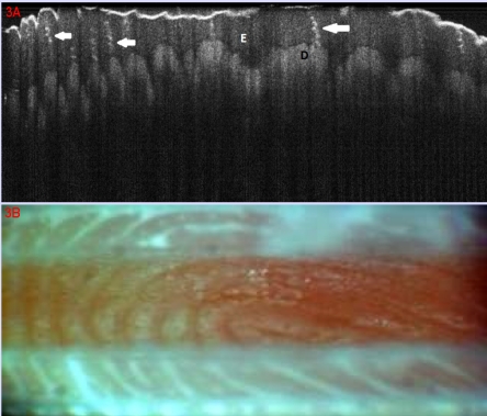
Methods: Preliminary results are presented from an initiated study. Fifteen consecutive patients with clinical suspicion of epithelial cancers and precancers registered over one week in a university dermatologic department were included. As control were selected 7 patients with inflammatory skin diseases (psoriasis, lichen planus, cutaneous lupus erythematosus). In all study and control patients, the lesions and samples of normal, perilesional skin were documented by clinical digital photography, contact dermoscopy with digital image capture and OCT with central wavelength of 930 nm. Final diagnosis was certified by histopathological analysis.

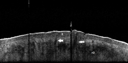
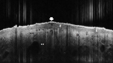
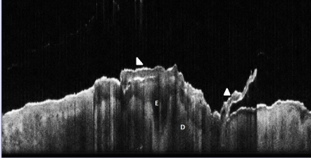
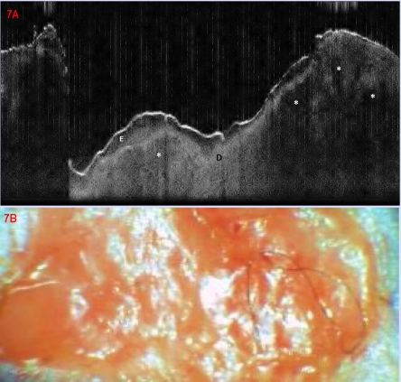
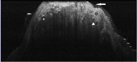
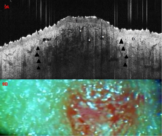
Results: We could identify morphological features in OCT examination that distinguished between normal and lesional skin, and between neoplastic vs. inflammatory lesions. In the same time, combining OCT and dermatoscopical evaluation of a lesion improved the performance of diagnosis when compared to clinical diagnosis alone and with either OCT or dermoscopy imaging used alone.

Conclusions: OCT appears as a promising method of in vivo diagnosis of early neoplastic cutaneous lesions with equivocal clinical and/or dermoscopic aspect. Continuation of our study as well as other larger investigation will be able to contribute with new insights in the role of OCT in the non-invasive diagnosis of skin disease.


Figures

Figure 1
Figure 1
Principle of function of Optical coherence
Figure 2
Figure 2
Normal skin, OCT aspect. Extensor aspect forearm, 53 year old male; E epidermis, D Dermis, rD reticular dermis, white arrows: hair follicles
Figure 3
Figure 3
Normal skin. Volar aspect of index finger; (A). OCT aspect: E epidermis, D dermis (dermal papillae), white arrows: sweat ducts; (B). Clinical aspect of dermatoglyphs
Figure 4
Figure 4
Normal skin, OCT aspect. Thigh, 53 years old female, E: epidermis, D: dermis, white arrows: blood vessels
Figure 5
Figure 5
Actinic Keratosis, OCT aspect. E: epidermis, slightly thickened in the lesional area; Arrowhead: signal–poor irregular band corresponding to hyperkeratosis; D dermis; asterisc black: round signal–free structures corresponding to dilated blood vessels; double asterisc white: venectasies
Figure 6
Figure 6
Bowen's Disease, OCT aspect. E: epidermis, which in the lesional area is markedly thickened, irregular, hypo–dense; one hair follicle appears to be involved; Hyperkeratosis is also marked, with a scale (arrowhead) partially detached. There is marked signal attenuation in the dermis (D)
Figure 7
Figure 7
Basal cell carcinoma. (A) OCT aspect, ; lobular signal–poor structure, corresponding to tumoral lobules; E epidermis, D Upper dermis; Upper dark band corresponds to keratin–rich stratum corneum; (B) Clinical aspect
Figure 8
Figure 8
Basal cell carcinoma, nodular type, OCT aspect. OCT aspect: ; signal–poor lobulated structures corresponding to tumoral lobules; D upper dermis; arrow: thin epidermis, difficult to distinguish from underlying dermis; arrowhead: round signal–free structures corresponding to cross–section blood vessels
Figure 9
Figure 9
Kaposi Sarcoma. (A) OCT aspect, focal changes in the central lesional area, showing a flattened epidermis (E), irregular, partly round partly branched and reticular signal–ppoor/free structures (asterisc) corresponding to vascular spaces, and a nodular area of attenuated signal (dark arrowheads) in the dermis (D); (B) clinical aspect of a small, superficial, red-bluish nodule on the knee of a 64 years old male
Figure 10
Figure 10
Seborrhoic keratosis. (A) OCT aspect E: epidermis, which in the lesional area, on the right half of the image shows marked thickening, with papillomatous aspect, as well as rounded hypo–dense structures (asteriscs), possibly corresponding to horn pseudocysts; marked signal attenuation over the dermis (D). (B) Clinical aspect of the margin area of a seborrhoic keratosis on the trunk
Figure 11
Figure 11
Psoriasis, OCT aspect. Epidermis (E) is hypo–dense and in the lesional area (left) is markedly thickened, with elongated protrusions in underlying dermis (D); overlying marked focal hyperkeratosis, as dark band, with scales protruding (arrowhead). There are longitudinal bands of signal attenuation corresponding to scaling
Figure 12
Figure 12
Cutaneous Sarcoidosis.(A) OCT aspect showing signal–poor agglomerated masses (asteriscs) occupying the dermis in the lesional area ( left), corresponding to granulomatous infiltrates. E epidermis; D dermis unaltered in the right extreme of the image, showing vascular spaces; (B) Clinical aspect of a well delimitated erythematous–brownish patch on the abdomen

Similar articles

Cited by

References

    1. Andersen P. Handbook of Non–Invasive Methods and the Skin. Florida: Boca Raton; 1996. Optical coherence tomography in dermatology.
    1. Welzel J. Optical coherence tomography in dermatology: a review. Skin Res Technol . 2001;7:1–9. - PubMed
    1. Tadrous PJ. Methods for imaging the structure and function of living tissues and cells: optical coherence tomography. J Pathol . 2000;(191):115–119. - PubMed
    1. Fujimoto JG. Optical biopsy and imaging using optical coherence tomography. Nat Med. 1995;1:970–972. - PubMed
    1. Welzel J, Lankenau E. Optical coherence tomography of the human skin . J Am Acad Dermatol. 1997;37:958–963. - PubMed

Publication types