. 2011 Feb;79(3):686-99.
doi: 10.1111/j.1365-2958.2010.07477.x. Epub 2010 Dec 7.

PAS/poly-HAMP signalling in Aer-2, a soluble haem-based sensor

Affiliations

PAS/poly-HAMP signalling in Aer-2, a soluble haem-based sensor

Kylie J Watts et al. Mol Microbiol. 2011 Feb.

Abstract

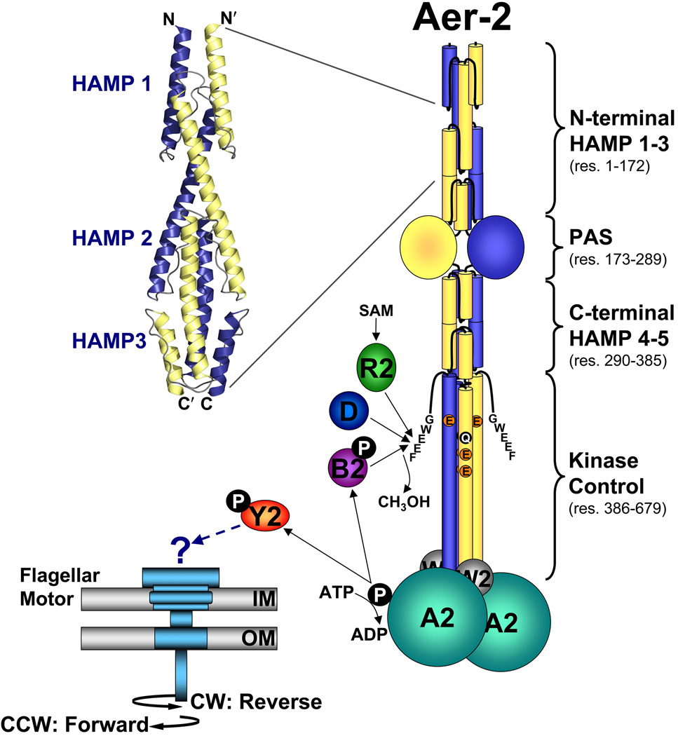
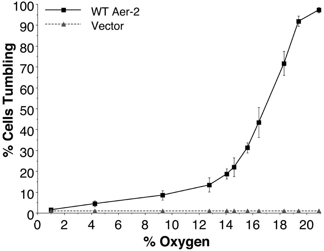
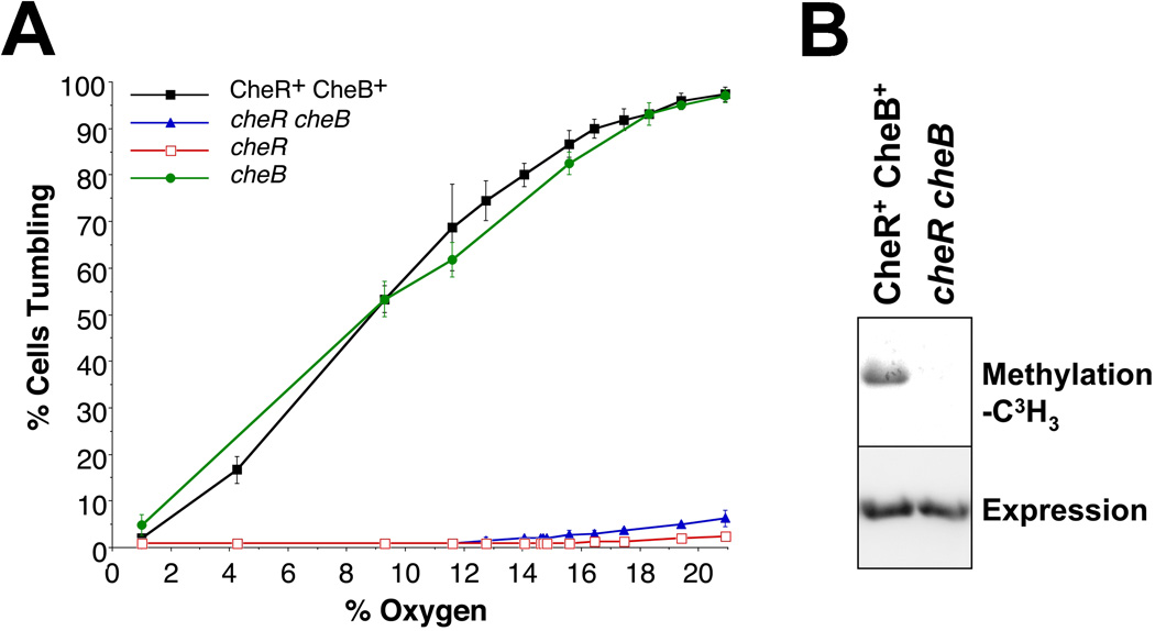
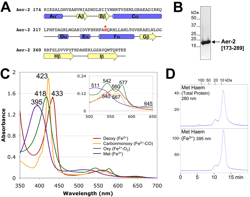
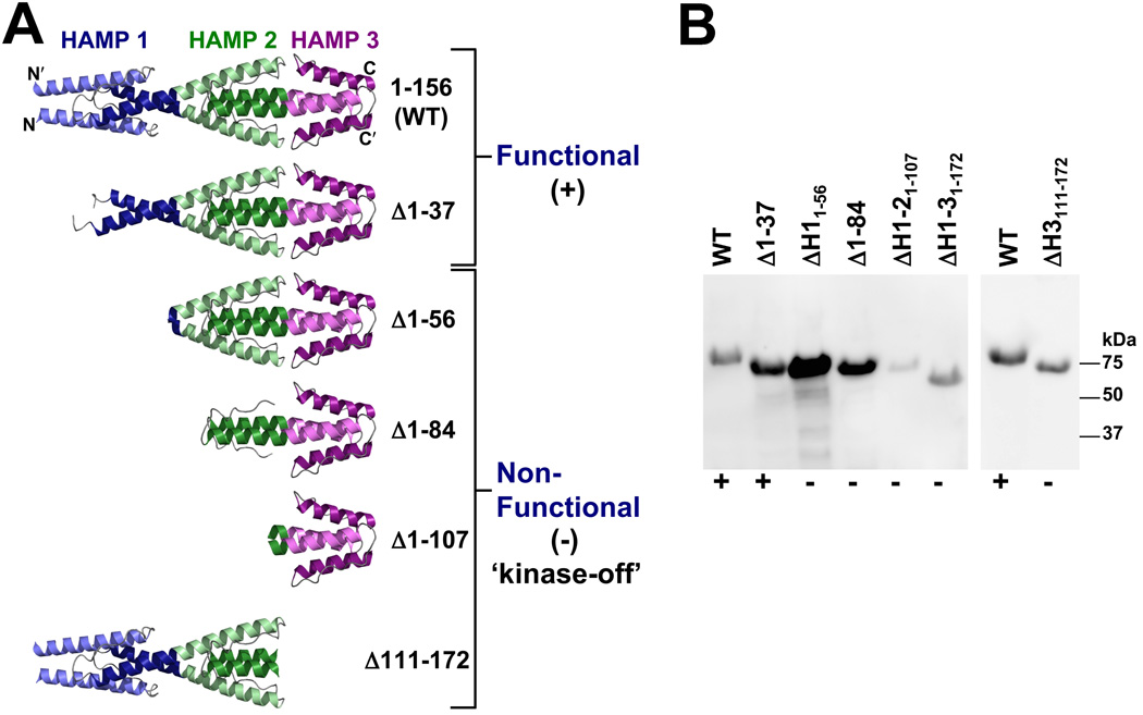
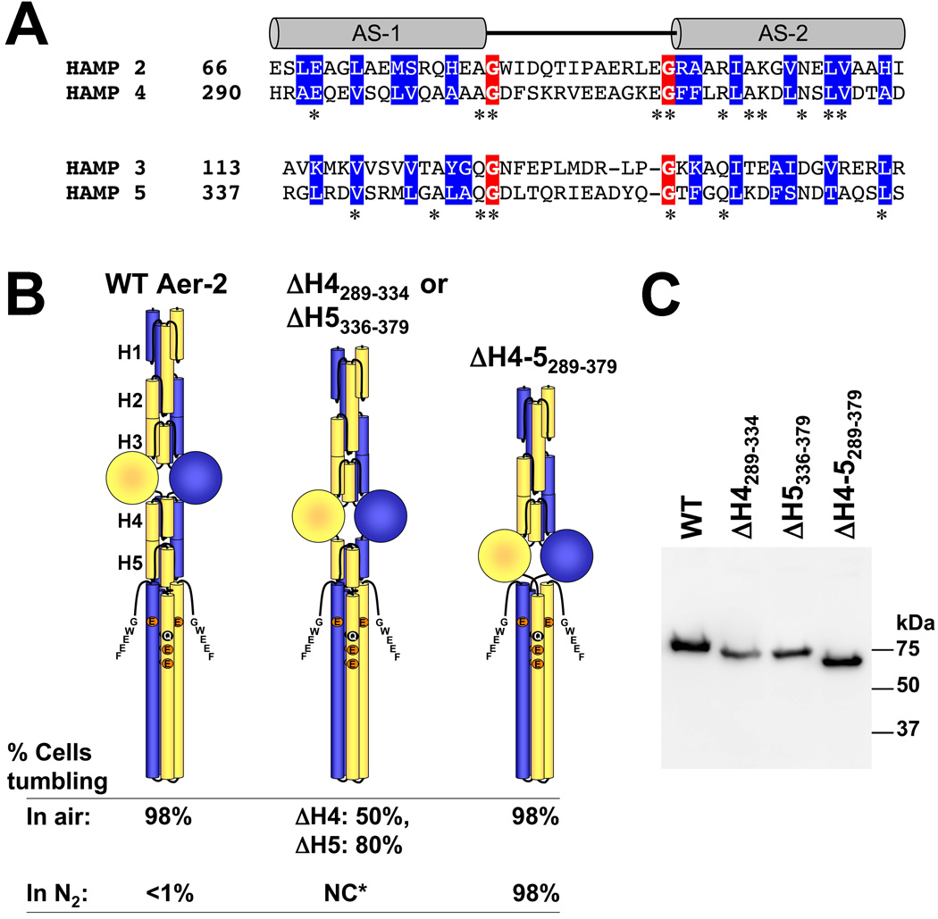
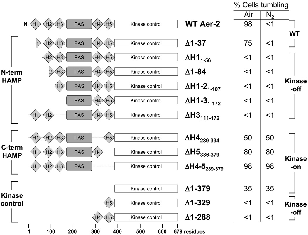
Poly-HAMP domains are widespread in bacterial chemoreceptors, but previous studies have focused on receptors with single HAMP domains. The Pseudomonas aeruginosa chemoreceptor, Aer-2, has an unusual domain architecture consisting of a PAS-sensing domain sandwiched between three N-terminal and two C-terminal HAMP domains, followed by a conserved kinase control module. The structure of the N-terminal HAMP domains was recently solved, making Aer-2 the first protein with resolved poly-HAMP structure. The role of Aer-2 in P. aeruginosa is unclear, but here we show that Aer-2 can interact with the chemotaxis system of Escherichia coli to mediate repellent responses to oxygen, carbon monoxide and nitric oxide. Using this model system to investigate signalling and poly-HAMP function, we determined that the Aer-2 PAS domain binds penta-co-ordinated b-type haem and that reversible signalling requires four of the five HAMP domains. Deleting HAMP 2 and/or 3 resulted in a kinase-off phenotype, whereas deleting HAMP 4 and/or 5 resulted in a kinase-on phenotype. Overall, these data support a model in which ligand-bound Aer-2 PAS and HAMP 2 and 3 act together to relieve inhibition of the kinase control module by HAMP 4 and 5, resulting in the kinase-on state of the Aer-2 receptor.


Figures

FIG. 1
FIG. 1
Cartoon showing the proposed domain organization of an Aer-2 dimer and the proteins associated with the Cluster II chemotaxis-like system. Each Aer-2 monomer contains three N-terminal HAMP domains, followed by a PAS sensing domain, two C-terminal HAMP domains, and a C-terminal kinase control domain. The structure of the three N-terminal HAMP domains was recently solved by x-ray crystallography [3LNR (Airola et al., 2010)]. By analogy to other methyl-accepting chemotaxis proteins, the kinase control domain is predicted to contain four methylation sites (QEEE, res. 414, 421, 428 and 610) and a C-terminal pentapeptide (GWEEF, res. 675–679) for binding the adaptation enzymes CheR2, CheB2 and CheD. Aer-2 is also predicted to form a ternary complex with the CheA2 and CheW2 proteins. The binding of an oxy-gas ligand to the Aer-2 PAS-heme domain is expected to alter the autophosphorylation rate of CheA2 and the subsequent transfer of phosphate to CheY2. It is currently unclear whether phospho-CheY2 binds to the flagellar motor or whether it interacts with a different response system. Abbreviations: A, W, Y, R, B, and D, Che proteins; SAM, S-adenosylmethionine; IM, inner membrane; OM, outer membrane; CW, clockwise; CCW, counterclockwise.
FIG. 2
FIG. 2
Steady-state behavior of receptor-less E. coli BT3388 cells expressing WT Aer-2 or vector alone, at decremental O2 concentrations between 20.9% and 1%. WT Aer-2 (pLH1) and the vector control (pProEX) were induced with 200 µM IPTG. Error bars represent the standard deviation from multiple titration experiments.
FIG. 3
FIG. 3
Aer-2-mediated behavior in E. coli in the presence or absence of the E. coli adaptation enzymes CheR and CheB. A. Steady-state behavior of isogenic E. coli strains (CheR+ CheB+ strain UU2612, cheR cheB strain UU2610, cheR strain UU2611, and cheB strain UU2632) expressing WT Aer-2 (with 200 µM IPTG) at O2 concentrations between 20.9% and 1%. Error bars represent the standard deviation from multiple titration experiments. B. Methylation of Aer-2 in the CheR+ CheB+ strain UU2612 and the absence of methylation in the cheR cheB strain UU2610 (upper panel), at similar levels of Aer-2 accumulation (lower panel, HisProbe Western blot).
FIG. 4
FIG. 4
Aer-2 PAS secondary structure, spectra and oligomeric state. A. Sequence and secondary structure of the Aer-2 PAS domain as predicted by PSIPRED (http://bioinf4.cs.ucl.ac.uk:3000/psipred) and a PAS sequence alignment created by L. Ulrich and I. Zhulin (personal communication). α-Helices and β-strands are shown as blue cylinders and yellow arrows, respectively. Aer-2 PAS contains a histidine in the Fα3 position (highlighted red), which coordinates heme in the structures of DOS and FixL. B. Coomassie-stained SDS-PAGE gel of purified Aer-2 PAS protein (res. 173–289, 16.3 kDa). C. Absorption spectra of purified Aer-2 PAS protein in the reduced (deoxy, dark red line), oxidized (met, purple line), carbonmonoxide-bound (carbonmonoxy, orange line) and oxygen-bound (oxy, green line) states. The absorbance maximum at each peak is indicated. The insert shows an expanded view of peaks between 500 and 650 nm. D. Elution profile of isolated Aer-2 PAS protein in its met-heme state during size-exclusion chromatography. The elution profile is shown in arbitrary units at 280 nm to reveal total protein content (top panel) and at 395 nm to detect the elution of met-heme (bottom panel). Fractions were removed and analyzed by Western blotting; in all cases, the Aer-2 PAS protein co-eluted with the heme (not shown).
FIG. 5
FIG. 5
Influence of N-terminal HAMP domains 1, 2 and 3 on Aer-2 signaling. A. Structure of the N-terminal HAMP domains (Airola et al., 2010) and models of the truncation mutants that were tested for function in behavioral assays. The Δ1–37 mutant was the only truncated product that retained Aer-2 function in E. coli (+). The other truncation mutants all resulted in kinase-off phenotypes (−). See text for details. B. HisProbe Western blot showing size differences among the truncated Aer-2 products as well as variations in their steady-state accumulation levels in E. coli BT3388 after induction with 50 µM IPTG. Abbreviation: ΔH, ΔHAMP.
FIG. 6
FIG. 6
Influence of C-terminal HAMP domains 4 and 5 on Aer-2 signaling. A. Sequence alignment of di-HAMP 2–3 and di-HAMP 4–5. Connector-flanking glycine residues are shaded red, whereas buried core residues (based on the resolved structure of HAMP 2–3) are shaded blue. Stars indicate identical N- and C-terminal HAMP residues. B. HAMP 4 and 5 are required to maintain the kinase-off state. Aer-2 deleted for HAMP 4 and 5 (far right) exhibited maximal kinase-on activity in air (tumbling in E. coli BT3388), like WT (far left). However, unlike WT, the ΔHAMP 4–5 mutant remained kinase-on (no change, NC) in N2 (in the absence of a diatomic oxy-gas). Mutant proteins lacking either HAMP 4 or HAMP 5 exhibited intermediate kinase-on activities (center figure). The HAMP 5 deletion began at residue 336 because residue 335 forms part of HAMP 4 and HAMP 5. C. HisProbe Western blot showing size differences among the internally truncated Aer-2 products and variations in their steady-state accumulation levels in E. coli BT3388 after induction with 50 µM IPTG. Abbreviations: AS, amphipathic sequence; H, HAMP; NC*, no change for either mutant.
FIG. 7
FIG. 7
Summary of Aer-2 deletions and the steady-state behavior mediated by these fragments in E. coli BT3388. Cells expressing WT Aer-2 (res. 1–679) or the Δ1–37 Aer-2 mutant tumbled in air and swam smoothly (<1% tumbling) in N2 (in the absence of a diatomic oxy-gas). Removing the N-terminal HAMP 2 and/or HAMP 3 domains resulted in a locked kinase-off phenotype, whereas removing C-terminal HAMP 4 and/or HAMP 5 resulted in a kinase-on phenotype. The default state of the isolated kinase control module was on, but it reverted to a kinase-off state in the presence of HAMP 5 or HAMP 4 and 5. All cells were induced with 200 µM IPTG for 45 min before monitoring their swimming behavior in a gas perfusion chamber. Mutants exhibiting less than 1% tumbling in air showed the same response after induction with 1 mM IPTG. Abbreviation: H, HAMP.

References

    1. Airola MV, Watts KJ, Bilwes AM, Crane BR. Structure of concatenated HAMP domains provides a mechanism for signal transduction. Structure. 2010;18:436–448. - PMC - PubMed
    1. Alexander RP, Zhulin IB. Evolutionary genomics reveals conserved structural determinants of signaling and adaptation in microbial chemoreceptors. Proc Natl Acad Sci U S A. 2007;104:2885–2890. - PMC - PubMed
    1. Aravind L, Ponting CP. The cytoplasmic helical linker domain of receptor histidine kinase and methyl-accepting proteins is common to many prokaryotic signalling proteins. FEMS Microbiol Lett. 1999;176:111–116. - PubMed
    1. Baraquet C, Theraulaz L, Iobbi-Nivol C, Mejean V, Jourlin-Castelli C. Unexpected chemoreceptors mediate energy taxis towards electron acceptors in Shewanella oneidensis. Mol Microbiol. 2009;73:278–290. - PubMed
    1. Bibikov SI, Biran R, Rudd KE, Parkinson JS. A signal transducer for aerotaxis in Escherichia coli. J Bacteriol. 1997;179:4075–4079. - PMC - PubMed

Publication types

MeSH terms