. 2011 Feb;79(3):814-25.
doi: 10.1111/j.1365-2958.2010.07490.x. Epub 2010 Dec 13.

Characterization of a new cytotoxin that contributes to Staphylococcus aureus pathogenesis

Affiliations

Characterization of a new cytotoxin that contributes to Staphylococcus aureus pathogenesis

Ashley L Dumont et al. Mol Microbiol. 2011 Feb.

Abstract

Staphylococcus aureus is an important pathogen that continues to be a significant global health threat because of the prevalence of methicillin-resistant S. aureus strains (MRSA). The pathogenesis of this organism is partly attributed to the production of a large repertoire of cytotoxins that target and kill innate immune cells, which provide the first line of defence against S. aureus infection. Here we demonstrate that leukocidin A/B (LukAB) is required and sufficient for the ability of S. aureus, including MRSA, to kill human neutrophils, macrophages and dendritic cells. LukAB targets the plasma membrane of host cells resulting in cellular swelling and subsequent cell death. We found that S. aureus lacking lukAB are severely impaired in their ability to kill phagocytes during bacteria-phagocyte interaction, which in turn renders the lukAB-negative staphylococci more susceptible to killing by neutrophils. Notably, we show that lukAB is expressed in vivo within abscesses in a murine infection model and that it contributes significantly to pathogenesis of MRSA in an animal host. Collectively, these results extend our understanding of how S. aureus avoids phagocyte-mediated clearance, and underscore LukAB as an important factor that contributes to staphylococcal pathogenesis.


Figures

Fig. 1
Fig. 1. LukAB is a potent staphylococcal cytotoxin that kills human phagocytic cell lines
A-B. Intoxication of Jurkat (A) or PMN-HL60 cells (B) with various dilutions of culture filtrate from the strain Newman (WT) and the indicated isogenic mutant strains. C. Abundance of toxins secreted by S. aureus strain Newman determined by LC-MS-MS and spectral counting. These data represent means of five independent experiments and the error bars represent the standard error of the mean (S.E.M.). D. Intoxication of PMN-HL60 cells with culture filtrate from WT containing an empty plasmid (WT/p), a strain lacking lukAB with an empty plasmid (ΔlukAB/p), and a strain lacking lukAB with a lukAB complementation plasmid (ΔlukAB/plukAB). E. Intoxication of PMN-HL60 cells with purified LukA (rLukA), LukB (rLukB), or a combination of rLukA and rLukB at the indicated concentrations. For the intoxications with both rLukA and rLukB, the total protein concentration is comprised of equal amounts of rLukA and rLukB, with each subunit at the indicated protein concentration. * indicates statistical significance from both rLukA and rLukB, P < 0.05. For panels (A, B and D), cell viability was monitored using CellTiter, where cells treated with medium were set at 100%. Results represent the average of triplicate samples ± standard deviation (S.D.). For panels (A and D), * indicates statistical significance from WT, ** indicates statistical significance from ΔlukAB/p, P < 0.05.
Fig. 2
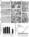
Fig. 2. LukAB disrupts the plasma membranes of target cells
A. Light microscopy images of PMN-HL60 cells or PMN-HL60 cells intoxicated for two hours with 5% (v/v) culture filtrate from the indicated S. aureus strain Newman (WT or the ΔlukAB strain). B. EM images of PMN-HL60 cells incubated in medium alone or PMN-HL60 cells intoxicated for one hour with 2.5% (v/v) culture filtrate from indicated strains. Swollen cellular morphology is indicted with an arrowhead. C. High magnification EM images of the plasma membrane of a PMN-HL60 cell intoxicated as described in panel B. Membrane disruption is indicated with an arrowhead. D. Intoxication of PMN-HL60 cells with culture filtrate from the indicated strain Newman isogenic mutants and the ΔlukAB/plukAB strain. * indicates statistical significance from WT, ** indicates statistical significance from ΔlukAB/p, P < 0.05. E. Intoxication of PMN-HL60 cells with purified LukA (rLukA), LukB (rLukB), or a combination of rLukA and rLukB at the indicated concentrations as described in Fig. 1. * indicates statistical significance from both rLukA and rLukB, P < 0.05. For panels (D and E) cells with compromised membranes were stained with SYTOX Green and green-fluorescence intensity was measured. Results represent the average of triplicate samples ± S.D.
Fig. 3
Fig. 3. LukAB kills primary human phagocytes
A. Intoxication of primary monocytes, macrophages, and dendritic cells (DC) with culture filtrate (2.5% v/v) from S. aureus strain Newman (WT) and the indicated isogenic mutant strains. Cell viability was monitored as described in Fig. 1. Results represent the mean from two donors, where cells from each donor were intoxicated with three independent exoprotein preparations, ± S.E.M. B. Intoxication of primary human PMNs with various dilutions of culture filtrates from the S. aureus strain Newman (WT) and the indicated isogenic mutant strains. Cell viability was monitored as described in Fig. 1. Results represent the mean from PMNs isolated from four donors ± S.E.M. C. Intoxication of primary human PMNs with culture filtrates (2.5% v/v) from the Newman WT/p, ΔlukAB/p, and ΔlukAB/plukAB strains. LDH-release was measured as an indicator of membrane disruption. Results represent the mean from PMNs isolated from six different donors ± S.E.M. D. Intoxication of primary human PMNs with purified rLukA, rLukB, or a combination of rLukA and rLukB at the indicated concentrations as described in Fig. 1. * indicates statistical significance from both rLukA and rLukB, P < 0.05. For panels (A-C) * indicates statistical significance from WT, ** indicates statistical significance from ΔlukAB/p, P < 0.05.
Fig. 4
Fig. 4. S. aureus lukAB mutants are attenuated in ex vivo infection models
A. Infection of PMN-HL60 cells with the S. aureus strain Newman WT and the indicated isogenic mutants at various MOI. Results represent the average of triplicate samples ± S.D. B. Infection of primary human PMNs with the S. aureus strain Newman WT, the lukAB isogenic mutant strain and complemented strain (ΔlukAB/plukAB) at various MOI. Results represent the mean from PMNs isolated from five donors ± S.E.M. C. Viability of the indicated S. aureus strains upon human whole blood infection and upon infection of primary human PMNs. Colony forming units were normalized where input S. aureus strains were set at 100% viable. Results represent the mean from whole blood isolated from six donors or the mean from PMNs isolated from 12 donors ± S.E.M. For panels (A and B) mammalian cells with compromised membranes were stained with SYTOX Green as described in Fig. 2. For panels (A-C) * indicates statistical significance from WT, ** indicates statistical significance from ΔlukAB/p, P < 0.05.
Fig. 5
Fig. 5. LukAB contributes to the cytotoxicity of MRSA and is important for the pathogenesis of MRSA in vivo
A. LukA, LukB, LukF-PV and α-toxin (Hla) production and the cytotoxic profiles of several MSSA and MRSA WT and isogenic ΔlukAB mutant strains against PMN-HL60 cells. PMN-HL60s were intoxicated with 10% (v/v) culture filtrate from the indicated strains and cell viability was monitored as described in Fig. 1. Results represent the average of triplicate samples ± S.D. B. Intoxication of primary human PMNs and primary human macrophages with culture filtrates (2.5% v/v) from the MRSA strain LAC, and the respective ΔlukAB isogenic mutant strains and complemented strains (ΔlukAB/plukAB). Cell viability was monitored as described in Fig. 1. Results represent the mean from PMNs and macrophages isolated from three to four donors ± S.E.M. C. Infection of primary human PMNs for one hour with the MRSA strain LAC WT, the lukAB isogenic mutant strain and complemented strain (ΔlukAB/plukAB) at various MOI. Results represent the mean from PMNs isolated from five donors ± S.E.M. D. Bioluminescent images of kidneys from mice infected with the WT S. aureus strain LAC containing pXen1 or the plukAB.Xen1. The kidneys of two representative mice per group are shown. E. Bacterial load recovered from the kidneys of mice infected retro-orbitally with the indicated S. aureus LAC strains. Each data point represents the number of bacteria (CFU) per milliliter of tissue homogenate in a single animal. Dashed line indicates the limit of detection. For panels (A-C and E) * indicates statistical significance from WT, ** indicates statistical significance from ΔlukAB/p, P < 0.05.

References

    1. Baba T, Bae T, Schneewind O, Takeuchi F, Hiramatsu K. Genome sequence of Staphylococcus aureus strain Newman and comparative analysis of staphylococcal genomes: polymorphism and evolution of two major pathogenicity islands. J Bacteriol. 2008;190:300–310. - PMC - PubMed
    1. Baba T, Takeuchi F, Kuroda M, Yuzawa H, Aoki K, Oguchi A, Nagai Y, Iwama N, Asano K, Naimi T, Kuroda H, Cui L, Yamamoto K, Hiramatsu K. Genome and virulence determinants of high virulence community-acquired MRSA. Lancet. 2002;359:1819–1827. - PubMed
    1. Bae T, Banger AK, Wallace A, Glass EM, Aslund F, Schneewind O, Missiakas DM. Staphylococcus aureus virulence genes identified by bursa aurealis mutagenesis and nematode killing. Proc Natl Acad Sci U S A. 2004;101:12312–12317. - PMC - PubMed
    1. Bae T, Schneewind O. Allelic replacement in Staphylococcus aureus with inducible counter-selection. Plasmid. 2006;55:58–63. - PubMed
    1. Brown EL, Dumitrescu O, Thomas D, Badiou C, Koers EM, Choudhury P, Vazquez V, Etienne J, Lina G, Vandenesch F, Bowden MG. The Panton-Valentine leukocidin vaccine protects mice against lung and skin infections caused by Staphylococcus aureus USA300. Clin Microbiol Infect. 2009;15:156–164. - PMC - PubMed

Publication types

LinkOut - more resources