. 2011 Jan 21;6(1):8.
doi: 10.1186/1750-1326-6-8.

Glutathione Peroxidase 4 is associated with Neuromelanin in Substantia Nigra and Dystrophic Axons in Putamen of Parkinson's brain

Affiliations

Glutathione Peroxidase 4 is associated with Neuromelanin in Substantia Nigra and Dystrophic Axons in Putamen of Parkinson's brain

Frederick P Bellinger et al. Mol Neurodegener. .

Abstract

Background: Parkinson's disease is a neurodegenerative disorder characterized pathologically by the loss of nigrostriatal dopamine neurons that project from the substantia nigra in the midbrain to the putamen and caudate nuclei, leading to the clinical features of bradykinesia, rigidity, and rest tremor. Oxidative stress from oxidized dopamine and related compounds may contribute to the degeneration characteristic of this disease.

Results: To investigate a possible role of the phospholipid hydroperoxidase glutathione peroxidase 4 (GPX4) in protection from oxidative stress, we investigated GPX4 expression in postmortem human brain tissue from individuals with and without Parkinson's disease. In both control and Parkinson's samples, GPX4 was found in dopaminergic nigral neurons colocalized with neuromelanin. Overall GPX4 was significantly reduced in substantia nigra in Parkinson's vs. control subjects, but was increased relative to the cell density of surviving nigral cells. In putamen, GPX4 was concentrated within dystrophic dopaminergic axons in Parkinson's subjects, although overall levels of GPX4 were not significantly different compared to control putamen.

Conclusions: This study demonstrates an up-regulation of GPX4 in neurons of substantia nigra and association of this protein with dystrophic axons in striatum of Parkinson's brain, indicating a possible neuroprotective role. Additionally, our findings suggest this enzyme may contribute to the production of neuromelanin.


Figures

Figure 1
Figure 1
GPX4 is associated with neuromelanin in SN neurons. GPX4 colocalizes with neuromelanin in SN of control brain. A. Western blot showing antibody specificity in human brain (left) and human and mouse neuroblastoma cells and mouse cortex (right). For "blocked" controls, antibody was preabsorbed with original recombinant antigen. B. Immunohistochemistry negative control with primary antibody omitted. Brown pigment is endogenous neuromelanin (black arrows). C. Robust GPX4 expression (dark grey Ni-DAB, white arrowheads) is associated with neuromelanin (brown, black arrows) and in the cytoplasm (*). D. Enlarged cell images, counterstained with methyl green to show nuclei of these neurons (white arrows). Scale bars: B, C, 20 μm, D, 10 μm.
Figure 2
Figure 2
GPX4 is found primarily in Neuromelanin-expressing neurons of SN. GPX4 immunoreactivity is present in most cells expressing neuromelanin but is absent in many cells expressing TH but not neuromelanin. A. Example of cells expressing TH but not neuromelanin. Light microscope images (above left) were filtered for neuromelanin (blue, above center) to compare with TH immunoreactivity (magenta, above right) and GPX4 immunoreactivity (green, below left). GPX4 images combined with neuromelanin (below, middle) and TH immunoreactivity (below, right) are also shown for comparison. GPX4 is present in surrounding glia but absent in TH-expressing neurons. B. Examples of TH and neuromelanin expressing neurons. GPX4 is present in most cells with neuromelanin. C. GPX4 immunoreactivity is prominent in neurons containing neuromelanin but no longer expressing TH. (Note: as images were not taken with confocal microscopy, color changes only show only co-localization of area and not intracellular co-localization). Scale bars: 20 μm.
Figure 3
Figure 3
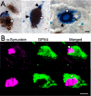
GPX4 in nigral inclusions of PD brain. A. GPX4 expression (dark grey Ni-DAB, white arrowheads) coincides with AS-positive Lewy bodies (blue BCIP, marked by black arrowheads) in SN. Black arrows indicate neuromelanin. B. Confocal microscope images of two cells showing relationship of GPX4 (green) to AS (magenta). The above example shows some co-localization of GPX4 with AS within a Lewy body, while the below example shows GPX4 only around the Lewy body perimeter. Scale bars: A, 20 μm, B, 10 μm.
Figure 4
Figure 4
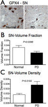
GPX4 is reduced overall but increased relative to cell density in PD SN. A. GPX4 (dark grey) is visibly reduced in SN of PD subjects (n = 12) compared to controls (n = 11). B. Total immunoreactivity of GPX4 is significantly reduced in SN of PD subjects compared to controls (P = 0.0398, Student's t-test). C. GPX4 immunoreactivity is increased relative to cell density (P = 0.0192, Student's t-test). Scale bars: 20 μm.
Figure 5
Figure 5
GPX4 expression in putamen. A. GPX4 immunoreactivity is detectable in cell bodies in the putamen of control brains (white arrowheads). B. Comparison of GPX4 labeling in normal and PD putamen. C. Mean GPX4 volume fraction is higher in PD (n = 12) putamen compared to control putamen (n = 11), although the difference is not significant (P > 0.05). D. Images of TH (Nova Red, red color, marked by black arrows) and GPX4 (DAB-Ni, blue/gray, white arrowheads) in putamen of non-PD (left) and PD (right) brain. Occasional swollen, dystrophic axons are present among the relatively few TH-positive fibers and terminals in PD putamen. Scale bars: 20 μm.
Figure 6
Figure 6
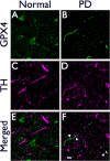
GPX4 is associated with dystrophic axons in PD putamen. Confocal images of GPX4 (blue) and TH (magenta) in non-PD (and PD brain (F, H, J). GPX4 has little association with TH in control brain sections, but colocalizes with TH in dystrophic axons in PD brain (shown by white color, white arrowheads). Scale bar: 10 μm.
Figure 7
Figure 7
Possible Role of GPX4 in Prevention of Apoptotic Cell Death and Production of Neuromelanin. In healthy DA neurons, GPX4 functions to reduce oxidized lipids by oxidizing glutathione (GSH) to glutathione disulfide (GSSG) (above). However, a buildup of oxidized dopamine metabolites or a reduction of glutathione may lead to the synthesis of neuromelanin by GPX4, a process that could compete with reduction of oxidized lipids (below). The accumulated oxidized lipids will be metabolized by 12/15-lipoxygenase (12/15-lipoxy) and may eventually induce AIF and promote apoptotic cell death of DA neurons.

References

    1. Adams JD Jr, Chang ML, Klaidman L. Parkinson's disease--redox mechanisms. Curr Med Chem. 2001;8:809–814. - PubMed
    1. Chinta SJ, Andersen JK. Dopaminergic neurons. Int J Biochem Cell Biol. 2005;37:942–946. doi: 10.1016/j.biocel.2004.09.009. - DOI - PubMed
    1. Halliday GM, Ophof A, Broe M, Jensen PH, Kettle E, Fedorow H, Cartwright MI, Griffiths FM, Shepherd CE, Double KL. Alpha-synuclein redistributes to neuromelanin lipid in the substantia nigra early in Parkinson's disease. Brain. 2005;128:2654–2664. doi: 10.1093/brain/awh584. - DOI - PubMed
    1. McCormack AL, Di Monte DA, Delfani K, Irwin I, DeLanney LE, Langston WJ, Janson AM. Aging of the nigrostriatal system in the squirrel monkey. J Comp Neurol. 2004;471:387–395. doi: 10.1002/cne.20036. - DOI - PubMed
    1. De Iuliis A, Burlina AP, Boschetto R, Zambenedetti P, Arslan P, Galzigna L. Increased dopamine peroxidation in postmortem Parkinsonian brain. Biochim Biophys Acta. 2002;1573:63–67. - PubMed

LinkOut - more resources