. 2011 Jan 21;8(1):6.
doi: 10.1186/1742-4690-8-6.

A nuclear export signal within the structural Gag protein is required for prototype foamy virus replication

Affiliations

A nuclear export signal within the structural Gag protein is required for prototype foamy virus replication

Noémie Renault et al. Retrovirology. .

Abstract

Background: The Gag polyproteins play distinct roles during the replication cycle of retroviruses, hijacking many cellular machineries to fulfill them. In the case of the prototype foamy virus (PFV), Gag structural proteins undergo transient nuclear trafficking after their synthesis, returning back to the cytoplasm for capsid assembly and virus egress. The functional role of this nuclear stage as well as the molecular mechanism(s) responsible for Gag nuclear export are not understood.

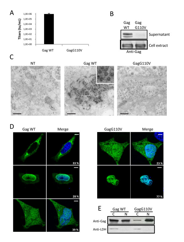
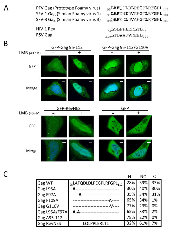
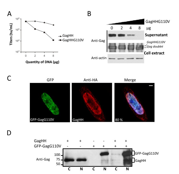
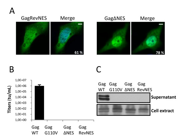
Results: We have identified a leptomycin B (LMB)-sensitive nuclear export sequence (NES) within the N-terminus of PFV Gag that is absolutely required for the completion of late stages of virus replication. Point mutations of conserved residues within this motif lead to nuclear redistribution of Gag, preventing subsequent virus egress. We have shown that a NES-defective PFV Gag acts as a dominant negative mutant by sequestrating its wild-type counterpart in the nucleus. Trans-complementation experiments with the heterologous NES of HIV-1 Rev allow the cytoplasmic redistribution of FV Gag, but fail to restore infectivity.

Conclusions: PFV Gag-Gag interactions are finely tuned in the cytoplasm to regulate their functions, capsid assembly, and virus release. In the nucleus, we have shown Gag-Gag interactions which could be involved in the nuclear export of Gag and viral RNA. We propose that nuclear export of unspliced and partially spliced PFV RNAs relies on two complementary mechanisms, which take place successively during the replication cycle.


Figures

Figure 1
Figure 1
Characterization of the GagG110V mutant. (A) Transduction rate of viruses harboring either GagWT or GagG110V. 293T cells were transfected for 48 h with FV vector encoding for GFP together with plasmids expressing Env, Pol and GagWT or GagG110V. Cell free supernatants were used to transduce 293T cells and the viral titer was determined from the number of GFP-positive cells by FACS analysis 48 h post-transduction. No infectivity was detected in the supernatant of GagG110V transfected cells, as observed in five independent experiments. (B) Western blotting performed on 293T cellular extracts and cell free supernatants shows the absence of viral particles in the supernatant of GagG110V transfected cells whereas intracellular GagG110V is normally produced. (C) Electron microscopy revealed, furthermore, the absence of intracellular capsids in 293T cells transfected with GagG110V. Bar: 0.5 μm. (D) Subcellular localization of GagWT and GagG110V in Hela transfected cells with GagWT or GagG110V and analyzed, 24 h post-transfection, by confocal microscopy following indirect immunofluorescence using rabbit polyclonal anti-PFV. GagWT is either nucleocytoplasmic, cytoplasmic or nuclear whereas GagG110V is mainly nuclear, as observed in three independent experiments (approximately 200 cells were counted in each preparation). (E) Western blotting performed on fractionated Hela cell extracts of Gag WT and GagG110V. Detection of the human lactate dehydrogenase (LDH) in cytoplasmic extracts only attests the validity of the fractionation assay (C: Cytoplasm, N: Nucleus).
Figure 2
Figure 2
Identification of a functional NES in PFV Gag. (A) Sequence alignment of a N-terminal region within Gag protein of primate foamy viruses. (B) Subcellular localization of GFP-Gag 95-112 and derived G110V mutant in Hela cells in the presence or the absence of LMB (40nM). GFP-RevNES and GFP alone were used respectively as positive and negative controls. Representative fluorescence images of the vast majority of cells expressing the indicated fusion proteins are shown by confocal microscopy. (C) Amino acid(s) important for Gag nuclear export. Point mutations or deletion were generated in the context of full length Gag and the resulting mutants were tested for sub-cellular localization after 24 h transfection using rabbit polyclonal anti-PFV antibodies. Results concerning Gag-RevNES localization were included. The numbers shown are the means of three independent experiments by counting 200 cells each (N: nuclear, NC: nucleocytoplasmic, C: cytoplasmic localization).
Figure 3
Figure 3
Dominant-negative properties of the GagG110V mutant. (A) Virus titers. Viral particles were produced in the supernatant of 293T cells transfected with the four-plasmid PFV vector system in the presence of increasing amounts of GagHH or GagHHG110V. Target 293T cells were transduced with cell free supernatants and titers were determined by FACS analysis 48 h post-transduction. Viral titers were dramatically reduced following addition of increasing amounts of GagHHG110V. This result is representative of three independent experiments. (B) Western blotting also shows a decrease in the amount of Gag proteins in supernatants whereas they are efficiently produced in 293T cells extracts. Therefore, GagG110V mutant negatively interferes with WT Gag impairing particles production. (C) Co-localization of GagHH and GFP-GagG110V. Hela cells were co-transfected with indicated plasmids and analyzed, 48 h post-transfection, by confocal microscopy following indirect immunofluorescence. GagWT colocalizes with GFP-GagG110V in the nucleus in 80 ± 4% of transfected cells in three independent experiments with approximately 100 cells counted each time. (D) Sequestration of GagWT by GagG110V in the nucleus. Nuclear interaction of GagHH and GFP-GagG110V revealed by co-immunoprecipitation of nuclear extracts of transfected Hela cells, using mouse anti-HA or anti-GFP antibodies followed by western-blotting performed with rabbit polyclonal anti-Gag antibodies (N : nucleus and C : cytoplasm).
Figure 4
Figure 4
HIV-1-RevNES fails to restore infectivity. (A) Subcellular localization of GagΔNES and Gag-RevNES in Hela cells analyzed 24h post-transfection with PFV antibodies by confocal microscopy following indirect immunofluorescence in three independent experiments (approximately 200 cells counted each time). (B) Transduction rate of viruses harboring either GagWT, GagG110V, GagΔNES or Gag-RevNES. Cell free supernatants were used to transduce 293T cells and the viral titer was determined from the number of GFP-positive cells by FACS analysis 48h post-transduction. No infectivity was detected in the supernatants of GagG110V, GagΔNES and Gag-RevNES transfected cells in four independent experiments. (C) Western blotting performed on 293T cell extracts and cell-free supernatants shows the absence of viral particles in the supernatants of GagG110V, GagΔNES and Gag-RevNES transfected cells whereas the intracellular Gag mutants are normally produced and matured.
Figure 5
Figure 5
Model for the possible nuclear role of FV Gag during the late stages of infection. (1) Full length viral RNA export is still unknown. (2) After synthesis in the cytoplasm, Gag protein enters the nucleus via its NLS domain (located within the GRII box). In the nucleus, Gag could interact with the full length viral RNA via its GRI box favoring Gag-Gag interaction and subsequently unmasking Gag NES. (3) The nuclear export factor, CRM1, also called exportin 1, would then be able to interact with this ribonucleoprotein complex leading to its efficient nuclear export. (4) In the cytoplasm, Gag proteins will multimerize for capsid assembly near the MTOC. In the absence of Gag proteins, the initial nuclear export of unspliced PFV RNA could rely on another export mechanism independent of these proteins.

References

    1. Freed EO. HIV-1 gag proteins: diverse functions in the virus life cycle. Virology. 1998;251:1–15. doi: 10.1006/viro.1998.9398. - DOI - PubMed
    1. Ganser-Pornillos BK, Yeager M, Sundquist WI. The structural biology of HIV assembly. Curr Opin Struct Biol. 2008;18:203–217. doi: 10.1016/j.sbi.2008.02.001. - DOI - PMC - PubMed
    1. Martin-Serrano J. The role of ubiquitin in retroviral egress. Traffic. 2007;8:1297–1303. doi: 10.1111/j.1600-0854.2007.00609.x. - DOI - PubMed
    1. Morita E, Sundquist WI. Retrovirus budding. Annu Rev Cell Dev Biol. 2004;20:395–425. doi: 10.1146/annurev.cellbio.20.010403.102350. - DOI - PubMed
    1. Klein KC, Reed JC, Lingappa JR. Intracellular destinies: degradation, targeting, assembly, and endocytosis of HIV Gag. AIDS Rev. 2007;9:150–161. - PubMed

Publication types

MeSH terms

Substances

LinkOut - more resources