

Review
doi: 10.1053/j.gastro.2011.01.027.
Epub 2011 Jan 18.
Targeted contrast-enhanced ultrasound: an emerging technology in abdominal and pelvic imaging
Affiliations
- PMID: 21255573
- PMCID: PMC4162392
- DOI: 10.1053/j.gastro.2011.01.027
Item in Clipboard
Review
Targeted contrast-enhanced ultrasound: an emerging technology in abdominal and pelvic imaging
Gastroenterology.
2011 Mar.
No abstract available
Conflict of interest statement
Conflicts of interest
The authors disclose no conflicts.
Figures
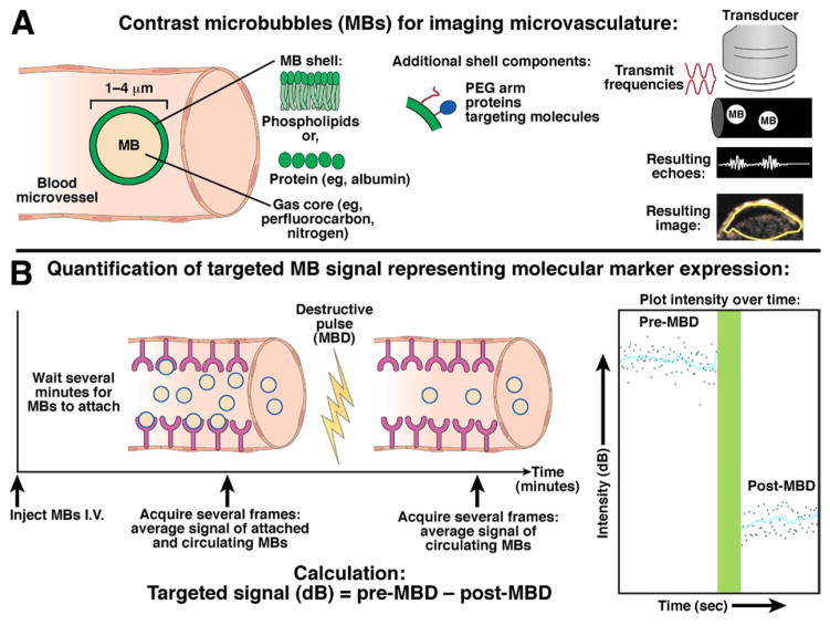
Principles of nontargeted and molecularly targeted contrast-enhanced ultrasound imaging with contrast microbubbles. (A) The gas core of lipid- or protein-shelled microbubbles (left) makes them highly echogenic compared to surrounding tissue and blood. Right, The pulse-inversion technique is commonly used for detection of microbubbles (see Supplementary Material). Two inverse-phase (red) pulses are transmitted through the tissue, and different echoes are reflected back from either microbubbles (MBs; white) or tissue (black). The resulting echo (summation from each pulse) from microbubbles (nonlinear behavior) is a distinct signature (white wave), whereas the waves reflected from the tissue (linear) signal cancel out (white flat line). The resulting ultrasound contrast-mode image shows a pixel-by-pixel distribution of the microbubble signal in a subcutaneous human colon cancer xenograft in a mouse. (B) Example of imaging sequence for quantification of molecular ultrasound imaging signal intensities within a region of interest. Please refer to text for more details.
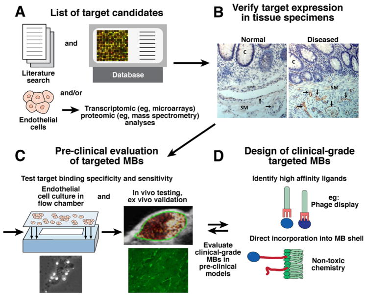
Possible approaches for molecular target discovery (step 1; A) and validation (step 2; B), testing of targeted contrast microbubbles (step 3; C), and clinical-grade contrast microbubble design (step 4; D). See text for more details. Example micrographs for target validation are immunohistochemically stained normal and diseased colon (C, crypt; SM, submucosa) tissues with hematoxylin-stained cell nuclei (blue) and target-stained (brown) vascular endothelial cells (black arrows); note that in this example, imaging targets are only expressed on vascular endothelial cells in diseased but not in normal tissue. Targeted microbubble can then be tested preclinically for binding specificity both in cell culture with flow chamber (schematic; brightfield micrograph [original magnification, ×100] shows white microbubbles attaching to cells) and in animal models in vivo in a subcutaneous human colon cancer xenograft (green outline) in a mouse. Target expression is also verified ex vivo by immunostaining; a 100-× micrograph shows brightly stained green blood vessels. Example of clinical-grade targeted microbubble design shows direct incorporation of binding ligand (eg, peptides identified by phage display; see also Supplementary Figure 1) into the microbubble shell.
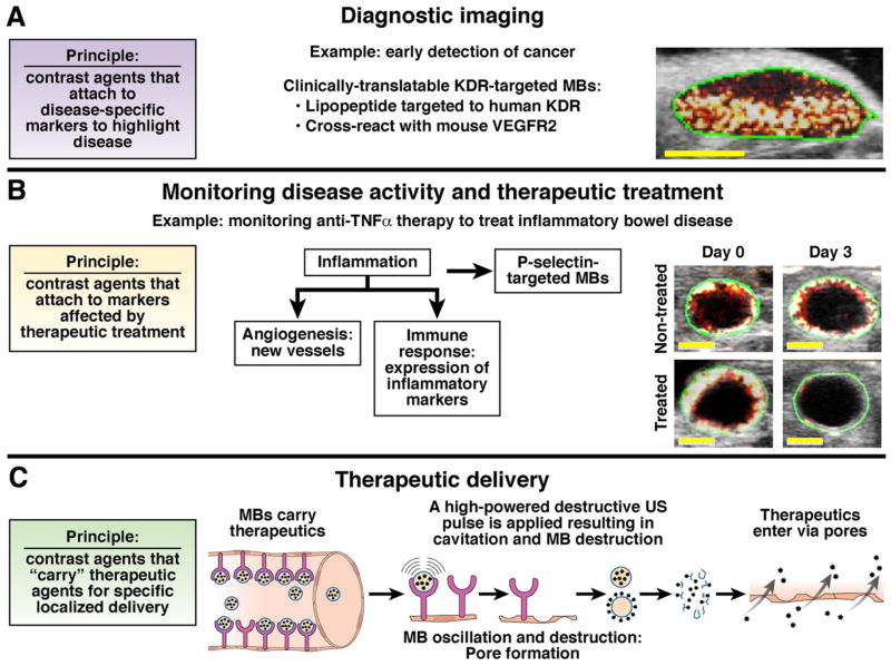
Nontargeted and molecularly targeted contrast-enhanced ultrasound imaging techniques can be used for several applications: Primary diagnostic imaging (detection and characterization of disease foci), monitoring disease activity and therapeutic treatment, and highly focused therapeutic delivery. (A) Early detection of cancer in a subcutaneous mouse xenograft by visualizing KDR, a marker of tumor angiogenesis expressed at early tumor stages (few mm of size; yellow bar, 3 mm), using KDR-targeted BR55 microbubbles. (B) Transverse ultrasound images show inflamed mouse colon (green region of interest around colon wall) visualized with contrast microbubbles (red and white colormetric map overlaid on B-mode image) targeted at inflammatory marker P-selectin, which is over-expressed in inflammatory bowel disease. (C) Nontargeted and/or disease-targeted (more focused delivery) microbubbles carrying therapeutics combined with ultrasound can be used to enhance therapeutic delivery to highly localized anatomical regions. Delivery process is described in text (also see Tinkov et al).
References
-
- Tardy I, Pochon S, Theraulaz M, et al. Ultrasound molecular imaging of VEGFR2 in a rat prostate tumor model using BR55. Invest Radiol. 2010;45:573–578. - PubMed
Publication types
MeSH terms
Substances
Grants and funding
LinkOut - more resources
Full Text Sources
Other Literature Sources
