. 2011 Feb 4;144(3):353-63.
doi: 10.1016/j.cell.2011.01.001. Epub 2011 Jan 20.

The RNA exosome targets the AID cytidine deaminase to both strands of transcribed duplex DNA substrates

Affiliations

The RNA exosome targets the AID cytidine deaminase to both strands of transcribed duplex DNA substrates

Uttiya Basu et al. Cell. .

Abstract

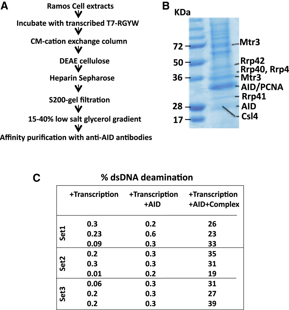
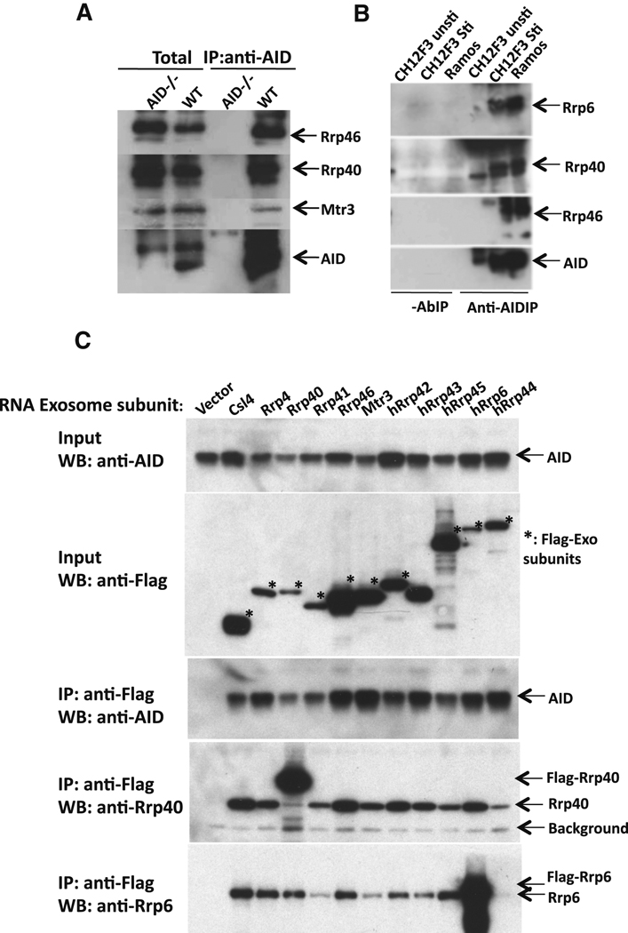
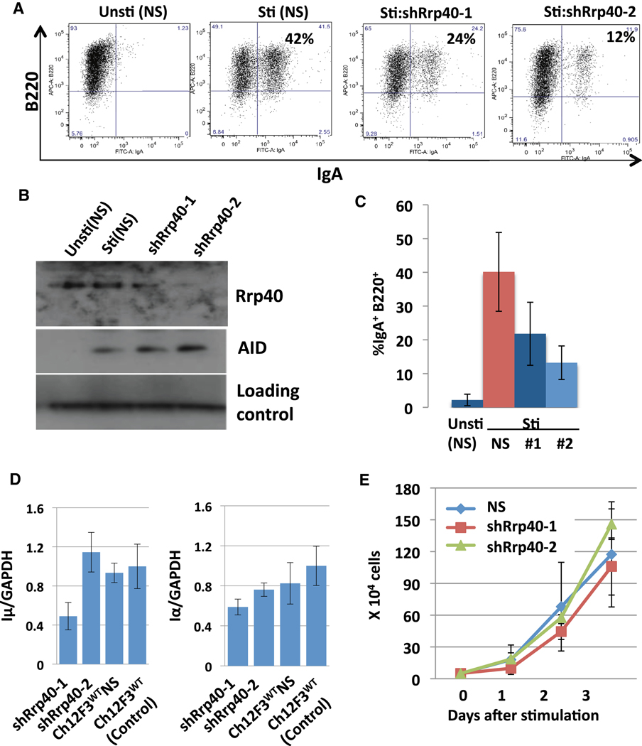
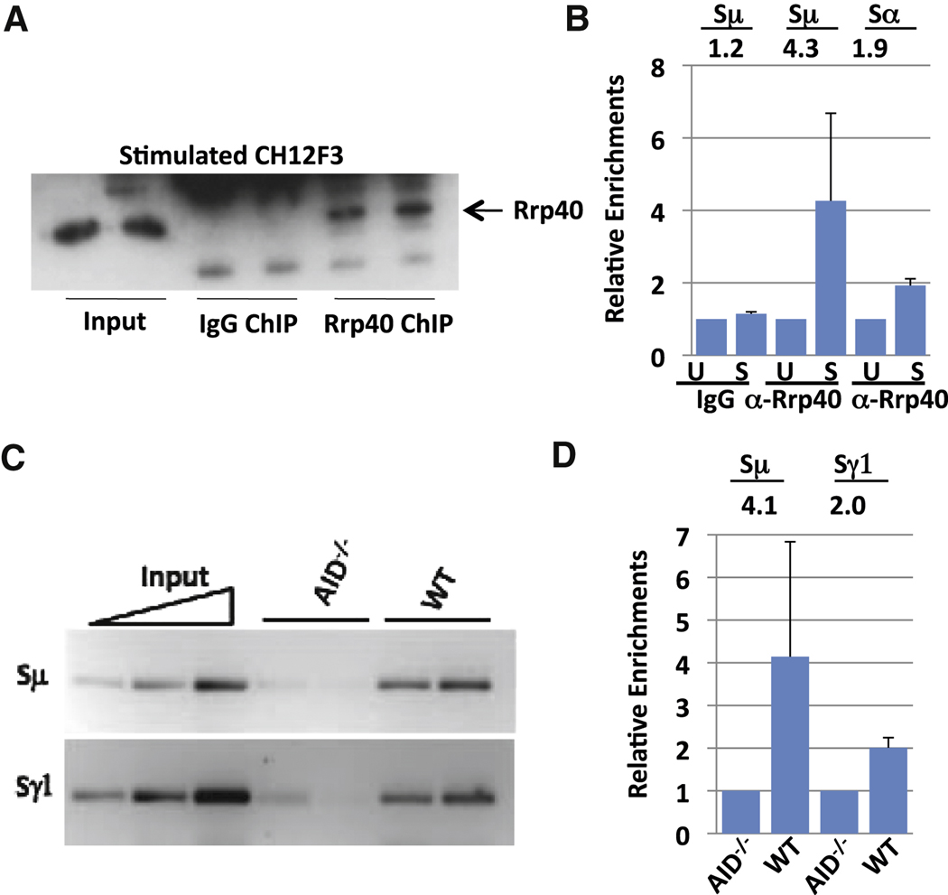
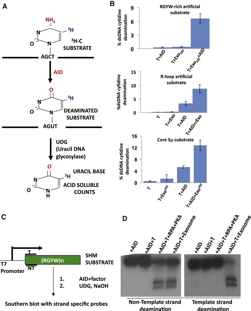
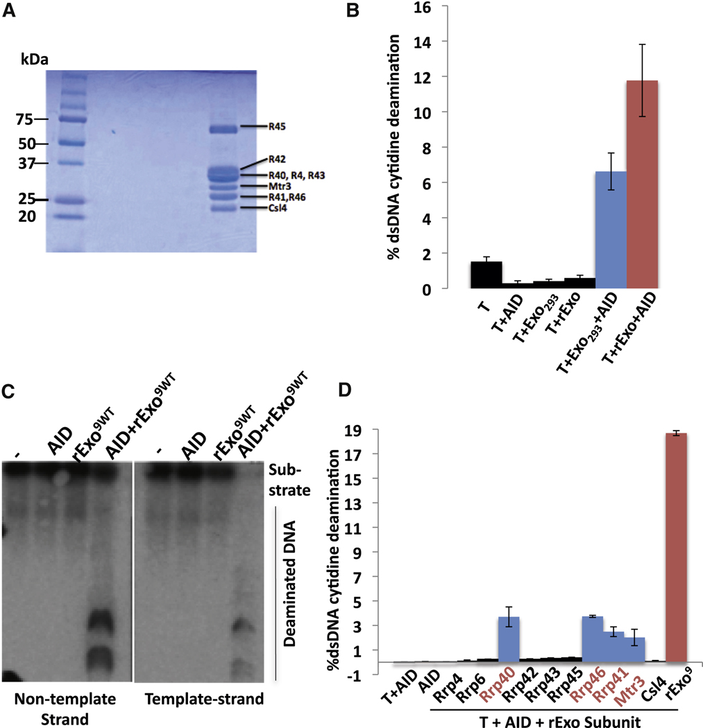
Activation-induced cytidine deaminase (AID) initiates immunoglobulin (Ig) heavy-chain (IgH) class switch recombination (CSR) and Ig variable region somatic hypermutation (SHM) in B lymphocytes by deaminating cytidines on template and nontemplate strands of transcribed DNA substrates. However, the mechanism of AID access to the template DNA strand, particularly when hybridized to a nascent RNA transcript, has been an enigma. We now implicate the RNA exosome, a cellular RNA-processing/degradation complex, in targeting AID to both DNA strands. In B lineage cells activated for CSR, the RNA exosome associates with AID, accumulates on IgH switch regions in an AID-dependent fashion, and is required for optimal CSR. Moreover, both the cellular RNA exosome complex and a recombinant RNA exosome core complex impart robust AID- and transcription-dependent DNA deamination of both strands of transcribed SHM substrates in vitro. Our findings reveal a role for noncoding RNA surveillance machinery in generating antibody diversity.


Figures

Figure 1
Figure 1. AID forms a transcription-Dependent complex with RNA Exosome
(A) Schematic outlining steps for enrichment of transcription dependent AID/RNA exosome/SHM substrate complex. Details are in text and supplementary methods. (B) Proteins enriched by purification scheme in panel A were analyzed by SDS-PAGE followed by staining with Coomassie Blue. Identity of proteins from selected bands was determined by mass spectrometry; bands that contain RNA exosome sub-units are indicted on the right with molecular weight markers on the left. (C) AID purified following ectopic expression in HEK293 cells was assayed in a 3H-release in vitro transcription dependent SHM substrate assay (Chaudhuri et al., 2003; see also Figure 5) in presence or absence of complex enriched by purification scheme in panel A. Percent of total transcribed DNA substrate deaminated is presented for 3 separate assays. See also Figure S1.
Figure 2
Figure 2. AID complexes with RNA exosome subunits in vivo
(A) AID immunoprecipitates from extracts of CSR-activated AID-deficient and wild-type B cells were assayed for Rrp40, Rrp46, Mtr3 and AID (indicated on the right) via Western blotting. The left two lanes show Western blotting of total extract and the right two lanes show Western blotting of immunoprecipitated products. (B) AID immunprecipitates ("Anti-AIDIP" lanes) or control reactions without anti-AID antibody ("-AbIP" lanes) from Ramos ("Ramos") and CSR-activated CH12F3 cells ("CH12F3 Sti") were assayed for Rrp46, Rrp40, Mtr3 and AID (indicated on right) by Western blotting. Un-stimulated CH12F3 cells ("CH12F3 unsti") were used as a negative control. (C) AID was co-expressed with individual FLAG-epitope tagged RNA exosome subunits in HEK293T cells. Lanes from left to right represent cells transfected with empty vector ("vector") as a control or the individual FLAG-epitope tagged subunit indicated at the top. The top two panels show Western blotting of total extract ("input") and the bottom three panels show western blotting with indicated antibodies (anti-AID, anti-Rrp40 and anti-Rrp6) following immunopreciptitation with anti-Flag antibodies. Asterisks indicate bands corresponding to Flag-tagged exosome subunits. A background band corresponding to the anti-Flag Ig light chain also is indicated (“Background").
Figure 3
Figure 3. RNA exosome subunit Rrp40 is required for normal CSR
(A) CH12F3 cells lenti-virally infected with a scrambled short hairpin plasmid (NS) or with shRNA against Rrp40 (shRrp40) were either not stimulated ("Unsti") or stimulated ("Sti") for 2 days with anti-CD40, IL4, and TGFβ and analyzed for IgA CSR by flow cytometry. ShRrp40-1 and shRrp40-2 are independent shRrp40-expressing CH12F3 isolates. Results are representative of 8 experiments; additional experiments shown in Fig. S2A,B. (B) NS, shRrp40-1 and shRrp40-2 expressing stimulated and un-stimulated CH12F3 isolates (shown in Panel A) were assayed for Rrp40 and AID by Western blotting. Results representative of 4 experiments; additional experiment in Fig. S2. (C) Average levels and standard deviation from the mean of CSR to IgA from three independent experiments (one shown in Panel A) performed simultaneously with un-stimulated ("unsti") NS, and stimulated ("Sti") NS, shRrp40-1 ("#1") and ShRrp40-2 ("#2") CH12F3 isolates. Five additional experiments gave similar results (Fig. S2A,B). (D) Total cellular RNA from 3 independently stimulated samples of indicated CH12F3 isolates (the ones used for Panel C) was assayed for Iµ transcripts (left panel) and Iα transcripts (right panel) via quantitative RT-PCR. Average and standard deviation from the mean is shown for the three separate experiments. Additional experiment based on Northern or RT-PCR is shown in Fig. S2F. (E) Growth curves of stimulated (NS), shRrp40-1 and shRrp40-2 CH12F3 cells calculated from three independent sets of three experiments (one used for panel C and others shown Fig. S2A) with a fourth set of 3 experiments indicated in Fig. S2E. Values represent average and standard deviation from the mean.
Figure 4
Figure 4. RNA exosome subunit Rrp40 is recruited to S regions
(A) Ch12F3 cells were either stimulated with TGFβ, IL4 and CD40 or kept un-stimulated for 48 hours. Subsequently, Rrp40 was immunoprecipitated from cell extracts under chromatin immunoprecipitation (ChIP) conditions and immunoprecipitates analyzed for Rrp40 by Western blotting with anti-Rrp40. (B) The "ChIPed" Rrp40-DNA complex from Ch12F3 cells was processed to isolate bound DNA. ChIPed DNA was tested for Sµ and Sα sequences via q-PCR. The average and standard deviation from the mean for three separate ChIP experiments is shown (see also Fig. S4). Numbers indicate average fold changes comparing stimulated and un-stimulated samples. Un-stimulated samples were arbitrarily normalized as 1 (Supp. Methods). (C) Rrp40 ChIPs were performed on extracts from primary splenic B cells stimulated for 2 days with anti-CD40 plus IL4. Sµ and Sγ1 were tested via semi-quantitative PCR; results are shown for two independent ChIP samples for each genotype. A 5-fold serial dilution of inputs is shown with the highest input concentration corresponding to 1/20 of total input. (D) ChIPed DNA from activated splenic B cells was tested for Sµ and Sγ1 via q-PCR. Numbers indicate average fold changes comparing WT and AID−/− samples. AID−/− samples were arbitrarily normalized as 1 (Supp. Methods). Values represent the average and standard deviation from the mean for three experiments. See Figure S4 for more details.
Figure 5
Figure 5. Cellular RNA exosome augments transcription-dependent AID deamination activity on template and non-template DNA strands
(A) Schematic representation of 3H release assay for AID deamination of transcribed dsDNA SHM substrate. (B) Top Panel: Results of 3H release assay in which a SHM substrate was transcribed by T7 polymerase (T) in the presence of purified AID (AID), purified HEK293 RNA exosome (Exo293) or both. Middle Panel: Results of 3H release assay in which a synthetic R-loop forming substrate was transcribed by T7 polymerase (T) in the presence of purified AID (AID), recombinant RNA exosome (rExo) or both. Bottom Panel: Results of 3H release assay in core Sµ substrate was transcribed by T7 polymerase (T) in the presence of purified AID (AID), purified HEK293 RNA exosome (Exo293) or both. In all three panels, values represent average and standard deviation from the mean from three independent experiments. (C) A schematic representation of assay for measuring strand-specific AID deamination of a transcribed dsDNA SHM. The location of template (T) and non-template (NT) strand probes is indicated. (D) The strand-specificity of RPA-dependent or RNA exosome-dependent DNA deamination was analyzed by the assay in panel C using either non-template (left panel) or template (right panel) strand-specific probes. Reactions contained AID, T7 polymerase (T), RPA and PKA, or purified HEK293 exosome ("Exosome") as indicated. See also Fig. S6.
Figure 6
Figure 6. Recombinant core RNA exosome and individual subunits stimulate AID activity
(A) Coomassie blue stained polyacrylamide gel electrophoreisis analysis of the 9 subunit recombinant RNA exosome complex generated from individual subunits. (B) Comparative analysis of the ability of RNA exosome complex from 293T cells (Exo293) and recombinant RNA core exosome complex (rExo) to stimulate AID deamination activity as measured by 3H release/SHM substrate assay in Fig. 5A. See Supp. Methods and Fig. S6 for details. Optimal amounts of RNA exosome293T and recombinant RNA exosome core complex were used (Fig. S6; Supp. Methods). (C) Assay of recombinant exosome complex to stimulate strand-specific AID deamination of a transcribed SHM substrate via assay in Fig. 5C. Non-template and template strand deamination are shown on left and right panels, respectively. Reactions contained either AID, recombinant core RNA exomsome (rExo9wt) or both (AID + rExo9wt) as indicated. (D) Individual recombinant RNA exosome subunits were assayed for ability to promote AID deamination of a T7-transcribed SHM substrate as outlined in Fig. 5A. Added exosome components are indicated. Control reactions with AID alone or AID plus T7 polymerase are on the left. A positive control with complete recombinant core RNA exosome (rExo9) plus T7 and AID is on the right. For panels B and D, values represent the average and standard deviation from the mean for three independent experiments. See also Figure S 6&7.

References

    1. Adelman K, Marr MT, Werner J, Saunders A, Ni Z, Andrulis ED, Lis JT. Efficient release from promoter-proximal stall sites requires transcript cleavage factor TFIIS. Mol Cell. 2005;17:103–112. - PubMed
    1. Anderson JR, Mukherjee D, Muthukumaraswamy K, Moraes KC, Wilusz CJ, Wilusz J. Sequence-specific RNA binding mediated by the RNase PH domain of components of the exosome. RNA. 2006;12:1810–1816. - PMC - PubMed
    1. Andrulis ED, Werner J, Nazarian A, Erdjument-Bromage H, Tempst P, Lis JT. The RNA processing exosome is linked to elongating RNA polymerase II in Drosophila. Nature. 2002;420:837–841. - PubMed
    1. Basu U, Chaudhuri J, Alpert C, Dutt S, Ranganath S, Li G, Schrum JP, Manis JP, Alt FW. The AID antibody diversification enzyme is regulated by protein kinase A phosphorylation. Nature. 2005;438:508–511. - PubMed
    1. Basu U, Wang Y, Alt FW. Evolution of phosphorylation-dependent regulation of activation-induced cytidine deaminase. Mol Cell. 2008;32:285–291. - PMC - PubMed

Publication types