. 2011 Feb 8;21(3):207-13.
doi: 10.1016/j.cub.2010.12.050. Epub 2011 Jan 20.

The Ndc80 loop region facilitates formation of kinetochore attachment to the dynamic microtubule plus end

Affiliations

The Ndc80 loop region facilitates formation of kinetochore attachment to the dynamic microtubule plus end

Jean-François Maure et al. Curr Biol. .

Abstract

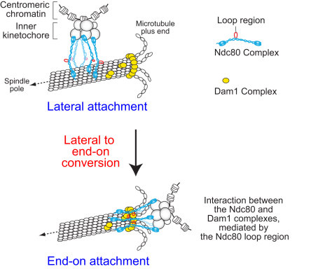
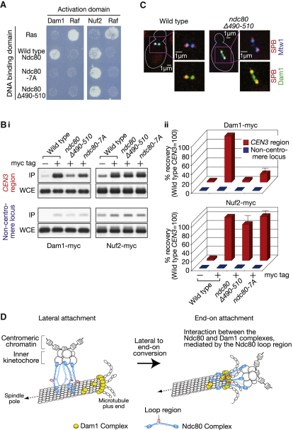
Proper chromosome segregation in mitosis relies on correct kinetochore-microtubule (KT-MT) interactions. The KT initially interacts with the lateral surface of a single MT (lateral attachment) extending from a spindle pole and is subsequently anchored at the plus end of the MT (end-on attachment). The conversion from lateral to end-on attachment is crucial because end-on attachment is more robust and thought to be necessary to sustain KT-MT attachment when tension is applied across sister KTs upon their biorientation. The mechanism for this conversion is still elusive. The Ndc80 complex is an essential component of the KT-MT interface, and here we studied a role of the Ndc80 loop region, a distinct motif looping out from the coiled-coil shaft of the complex, in Saccharomyces cerevisiae. With deletions or mutations of the loop region, the lateral KT-MT attachment occurred normally; however, subsequent conversion to end-on attachment was defective, leading to failure in sister KT biorientation. The Ndc80 loop region was required for Ndc80-Dam1 interaction and KT loading of the Dam1 complex, which in turn supported KT tethering to the dynamic MT plus end. The Ndc80 loop region, therefore, has an important role in the conversion from lateral to end-on attachment, a crucial maturation step of KT-MT interaction.


Figures

None
Graphical abstract
Figure 1
Figure 1
Deletions and Mutations within the Ndc80 Loop Region Cause Cell Lethality or Temperature-Sensitive Cell Growth (A) Step-wise development of kinetochore (KT)-microtubule (MT) interaction during prometaphase (i–v) and metaphase (vi). See more detail in [1]. (B) Structure of the Ndc80 complex, which consists of four proteins [8–10]. The position of the loop region is indicated. (C) The probability of forming coiled-coil motifs along amino acid residues of Ndc80 protein in Saccharomyces cerevisiae. Top: Full length of Ndc80. Bottom: Amino acid residues 420–550. Thick red lines indicate the positions of deletions in Ndc80 mutants, constructed in this study. ts, temperature-sensitive cell growth. (D) Multiple sequence alignment of the Ndc80 loop region from different organisms. The regions with coiled-coil probability <0.5 were selected for alignment. Some residues (numbers in parentheses) showed less conservation and were not shown here. Conserved residues are highlighted in colors: hydrophobic (light blue), acidic (purple), and basic (red) residues, asparagine (green), proline, and glycine (yellow). The positions of alanine substitution for the ndc80-7A mutant are shown in red rectangles. (E) ndc80Δ490-510 and ndc80-7A mutants show temperature-sensitive cell growth. 10-fold serial dilutions of wild-type (T6500), ndc80Δ490-510 (T6566), and ndc80-7A (T7881) cells were spotted onto YPD plates and incubated at the 25°C (top) and 35°C (bottom) for 48 hr. (F) Wild-type and ndc80Δ490-510, ndc80-7A cells show similar Ndc80 expression levels and similar Nuf2 association with Ndc80. NUF2-myc cells with NDC80 wild-type (T7082), ndc80Δ490-510 (T7085), or ndc80-7A (T8357), tagged with HA, were treated with α factor, released to fresh YPD medium at 35°C, and harvested after 70 min from the release (at which time the majority of cells were in metaphase). Wild-type cells without HA or myc tags (T7084, T6981) were also treated in the same way as controls. Total proteins (top) and the proteins immnunoprecipitated with a myc antibody (bottom) were detected by western blotting with an HA antibody.
Figure 2
Figure 2
In Mutants of the Ndc80 Loop Region, the Initial KT-MT Interaction Occurs Normally, but Sister KT Biorientation Is Established Inefficiently (A) The initial KT-MT interaction: wild-type (T7848), ndc80Δ490-510 (T7862), and ndc80-7A (T8397) cells with CEN5-tetOs TetR-3×CFP Venus-TUB1 were treated with α factor and released to fresh YPD medium at 35°C. CFP and Venus images were acquired every 10 s at 35°C. To avoid photo-bleaching of fluorescence signals during image acquisition, the field of observation was changed every 10 min. (i) Representative live-cell images, in which a wild-type cell showed CEN5 detachment from, and subsequent reattachment to, MTs. The cell shape is outlined in white. ndc80Δ490-510 and ndc80-7A mutants showed similar behavior of CEN5 (data not shown). (ii) Timing of CEN5 detachment from MTs, shown as the percentage of cells per field showing detachment during each 10 min time window. (iii) Duration of CEN5 dissociation from MTs in individual cells (means and standard errors). n.s., difference is not significant. (B) Establishment of sister KT biorientation. T7848, T7862, and T8397 cells (see A) were treated as in (A). CFP and Venus images were acquired every 1 min at 35°C. (i) Representative images of wild-type and ndc80-7A mutant cells, which showed separated and unseparated sister CEN5s, respectively. (ii) The percentage of cells showing separation of sister CEN5s on the bipolar spindle for at least two consecutive time points, until indicated time points (0 min: establishment of bipolar spindle). Sister CEN5s were scored as “separated” when two signals were discernible. See also Figure S2.
Figure 3
Figure 3
Mutants of the Ndc80 Loop Region Show Inefficient Conversion from Lateral to End-on KT-MT Attachment (A) Experimental system to analyze KT interaction with individual MTs. See details in [11]. (B) Mutants of the Ndc80 loop region show normal initial KT interaction with MTs, but subsequent establishment of sister KT biorientation is inefficient. NDC80 wild-type (T6803), ndc80Δ490-510 (T6690), and ndc80-7A (T7955) cells with PGAL-CEN3-tetOs TetR-GFP Venus-TUB1 PMET3-CDC20 were treated with α factor in methionine drop-out medium with 2% raffinose for 2.5 hr and released to YP medium containing 2% galactose, 2% raffinose, and 2 mM methionine at 25°C to inactivate CEN3 and arrest cells in metaphase. After 3 hr, the culture temperature was changed to 35°C. After 15 min, cells were suspended in the same medium but containing 2% glucose instead of galactose/raffinose to reactivate CEN3 (defined as 0 min). Cells were collected at indicated time points and fixed with paraformaldehyde. GFP and Venus images were acquired and CEN3-MT interaction was scored as indicated in the schematic drawing. In most of spc24-1 cells analyzed in this assay, CEN3 remained uncaptured by MTs for 60 min (Figure 2c in [11]), in contrast to the ndc80 loop-region mutants. (C) Mutants of the Ndc80 loop region show inefficient conversion from the lateral to end-on KT-MT attachment. T6803, T6690, and T7955 cells (see B) were treated as in (B), except that cells were suspended in synthetic complete medium containing 2% glucose and 2 mM methionine to reactivate CEN3. Cells were immobilized and GFP and Venus images were acquired every 20 s for 30 min at 35°C. (i) When the plus end of a shrinking MT caught up with CEN3, the MT subsequently showed either regrowth (MT rescue) or tethering of CEN3 to its plus end while shrinking further (end-on attachment and end-on pulling). Representative images of the events in wild-type cells and a graph showing frequency of the two events; these events happened in two mutants as in wild-type cells, albeit with very different frequencies. (ii) Frequency of each mode of CEN3 transport by a MT toward a spindle pole. Modes were classified as indicated by the schematic drawing. Sliding and end-on pulling were scored only when CEN3 moved for 1 μm or longer by each mode of the transport. The pink bars represent the cases where the end-on attachment was established before CEN3 moved along the MT lateral side more than 1 μm.
Figure 4
Figure 4
The Ndc80 Loop Region Is Required for Ndc80-Dam1 Interaction and for Dam1 Loading on the KT (A) The Ndc80 loop region is required for Ndc80-Dam1 interaction in a two-hybrid assay. The same amount of cells expressing indicated proteins, fused with a DNA binding domain or an activation domain, were spotted on histidine drop-out plates and incubated at 35°C for 48 hr. Cell growth suggests interaction between the two relevant proteins. Ras and Raf were used as controls for the assay. (B) The Ndc80 loop region is required for Dam1 loading on the KT. DAM1-myc cells with wild-type (T8761), ndc80Δ490-510 (T8762), and ndc80-7A (T8763) were treated with α factor, released to fresh YPD medium at 35°C, harvested after 70 min from the release (at which time the majority of cells were in metaphase), and treated with formaldehyde to crosslink. NUF2-myc cells with wild-type (T8777), ndc80Δ490-510 (T8778), and ndc80-7A (T8779) were treated in the same way. Wild-type cells without myc tags (T6500) were also treated in the same way, as a control. (i) Gel images of PCR products, amplified at CEN3 region and at a noncentromere locus (MPS1 locus, 45 kb from CEN4), with total DNA from whole cell extract (WCE) or immunoprecipitated DNA (IP) as a template. (ii) The percentage of recovered DNA was first quantified as a fraction of corresponding WCE in individual samples. Then, these percentage values were standardized, relative to that in NDC80 wild-type cells (at CEN3 region). Mean and standard errors were obtained from three independent experiments. (C) The Ndc80 loop region is required for Dam1 colocalization with the KT. Wild-type (T7868) and ndc80Δ490-510 (T7866) cells with DAM1-3×GFP MTW1-3×CFP SPC42-RFP were cultured and harvested as in (B). Representative images are shown here. Spindle pole bodies (SPBs) were visualized with Spc42-RFP. Other representative images and the quantification of total Dam1 and Mtw1 signals in individual cells are shown in Figure S4A. (D) Summary for the role of the Ndc80 loop region (shown in red) in the conversion of lateral to end-on KT-MT attachment. During lateral attachment, the Ndc80 complex (blue) binds a MT, presumably at its Ndc80/Nuf2 CH domains and the N-terminal region of Ndc80 [8–10]. To convert lateral attachment to end-on attachment, it is crucial that the Ndc80 loop region mediates the interaction with the Dam1 complex (yellow), which localizes at the MT plus end and forms an oligomer and/or a ring encircling the MT [3, 7, 21]. The Ndc80-Dam1 interaction could be direct or indirect, and more factors might be involved in this interaction. See also Figure S4.

Comment in

References

    1. Tanaka T.U. Kinetochore-microtubule interactions: Steps towards bi-orientation. EMBO J. 2010;29:4070–4082. - PMC - PubMed
    1. Grishchuk E.L., Molodtsov M.I., Ataullakhanov F.I., McIntosh J.R. Force production by disassembling microtubules. Nature. 2005;438:384–388. - PubMed
    1. Tanaka K., Kitamura E., Kitamura Y., Tanaka T.U. Molecular mechanisms of microtubule-dependent kinetochore transport toward spindle poles. J. Cell Biol. 2007;178:269–281. - PMC - PubMed
    1. Joglekar A.P., Bloom K.S., Salmon E.D. Mechanisms of force generation by end-on kinetochore-microtubule attachments. Curr. Opin. Cell Biol. 2010;22:57–67. - PMC - PubMed
    1. Santaguida S., Musacchio A. The life and miracles of kinetochores. EMBO J. 2009;28:2511–2531. - PMC - PubMed

Publication types

MeSH terms

Substances