. 2011 Feb 8;21(3):214-20.
doi: 10.1016/j.cub.2010.12.048. Epub 2011 Jan 20.

Ndc80 internal loop interacts with Dis1/TOG to ensure proper kinetochore-spindle attachment in fission yeast

Affiliations

Ndc80 internal loop interacts with Dis1/TOG to ensure proper kinetochore-spindle attachment in fission yeast

Kuo-Shun Hsu et al. Curr Biol. .

Abstract

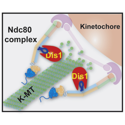
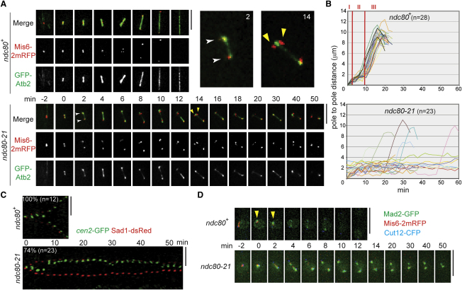
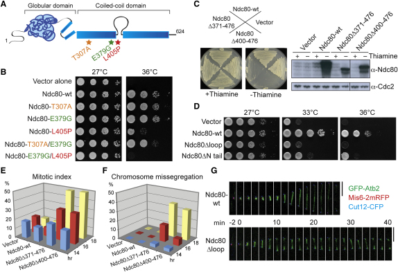
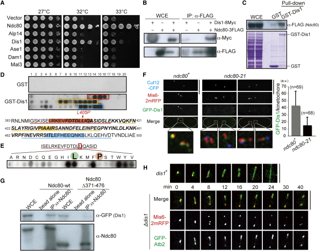
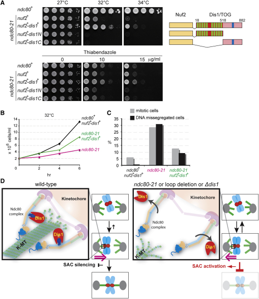
The Ndc80 complex, a conserved outer kinetochore complex, comprising four components (Ndc80/Hec1, Nuf2, Spc24, and Spc25), constitutes one of the core microtubule-binding sites within the kinetochore. Despite this knowledge, molecular mechanisms by which this complex contributes to establishment of correct bipolar attachment of the kinetochore to the spindle microtubule remain largely elusive. Here we show that the conserved internal loop of fission yeast Ndc80 directly binds the Dis1/TOG microtubule-associated protein, thereby coupling spindle microtubule dynamics with kinetochore capture. Ndc80 loop mutant proteins fail to recruit Dis1 to kinetochores, imposing unstable attachment and frequent spindle collapse. In these mutants, mitotic progression is halted attributable to spindle assembly checkpoint activation, and chromosomes remain in the vicinity of the spindle poles without congression. dis1 deletion precisely phenocopies the loop mutants. Intriguingly, forced targeting of Dis1 to the Ndc80 complex rescues loop mutant's defects. We propose that Ndc80 comprises two microtubule-interacting interfaces: the N-terminal region directly binds the microtubule lattice, while the internal loop interacts with the plus end of microtubules via Dis1/TOG. Therefore, our results provide a crucial insight into how the Ndc80 complex establishes stable bipolar attachment to the spindle microtubule.


Figures

None
Graphical abstract
Figure 1
Figure 1
The ndc80-21 Mutant Is Defective in Spindle-Kinetochore Attachment with Activation of the Spindle Assembly Checkpoint (A) Prolonged mitotic delay with unstable spindle microtubules. Time-lapse fluorescence montages of spindle microtubules (GFP-Atb2, α2-tubulin, green) and kinetochores (Mis6-2mRFP, kinetochore component Mis6 tagged with two copies of monomeric RFP, red) are shown (wild-type, top, and ndc80-21, bottom, cultured at 36°C). Merged images of two time points (2 min and 14 min) were enlarged in the top right corner. See Movie S1 (wild-type) and Movie S2 (ndc80-21). (B) Profiles of mitotic progression. Changes of the inter-SPB distance are plotted against time in wild-type (top) or ndc80-21 (lower). Phase I is a period of initial spindle elongation (I), while phase II represents the duration in which spindle length is constant (II) and phase III corresponds to anaphase B (III) [11]. (C) Time-lapse analysis of sister centromere behavior. Wild-type (top) or ndc80-21 (bottom) cells containing cen2-GFP (green) and Sad1-dsRed (red, an SPB marker) cultured at 36°C were recorded (2 min intervals) and converted to kymograph. See Movie S3 (wild-type) and Movie S4 (ndc80-21). (D) Mad2 localization at kinetochores. Wild-type (top) or ndc80-21 (bottom) cells containing Mad2-GFP (green), Mis6-2mRFP (kinetochore, red), and Cut12-CFP (SPB) was shifted to 36°C for 40 min and mitotic images were recorded (see Supplemental Experimental Procedures). Arrowheads show transient kinetochore localization (∼2 min) of Mad2 in wild-type. See Figures S1C and S1D for additional data. Scale bars represent 5 μm.
Figure 2
Figure 2
The Internal Loop Is Essential for Ndc80's Role in Spindle Microtubule Stabilization (A) A schematic view of Ndc80 with three mutation sites (asterisks) in ndc80-21. See Figures S2A and S2B for more information. (B) Identification of the mutation site(s) responsible of temperature sensitivity. The ndc80 gene containing indicated mutation(s) were introduced into an ndc80-21 strain as episomal plasmids (Table S2). 10-fold serial dilution (5 × 104 cells in the first spot) was applied on rich media, followed by incubation at 27°C or 36°C for 3 days. (C) Dominant-negative effects of overproduced loop-less Ndc80. Cells carrying indicated plasmids were streaked on minimal plates with (left plate, repressed) or without (right plate, derepressed) thiamine. For immunoblotting, 30 μg protein extracts were run on the gel. The Ndc80 protein was detected with Ndc80 antibody (Supplemental Experimental Procedures). (D) Nonfunctionality of loop-less Ndc80. Plasmids producing wild-type, loop-less (Ndc80Δ400-476, Ndc80Δloop), or N-terminally truncated (Ndc80Δ2-94, Ndc80ΔN) Ndc80 were introduced into an ndc80-21 strain. (E and F) Defective mitosis in cells overproducing loop-less Ndc80. Cells used in (C) were grown in minimal liquid media in the absence of thiamine, and the percentage of mitotic cells (E, microtubule morphology and the number of SPBs) and patterns of chromosome segregation (F, kinetochores and DAPI) were observed (n > 200). (G) Mitotic arrest accompanied with unstable spindle microtubules. Live image analysis was performed in cells overproducing Ndc80ΔN (16 hr in the absence of thiamine, bottom row). Scale bars represent 5 μm.
Figure 3
Figure 3
Dis1/TOG Microtubule-Associated Protein Is a Factor Responsible for Ndc80 Loop-Dependent Kinetochore Microtubule Stabilization (A) Rescue of ndc80-21 by multicopy plasmids containing dis1+. See Figure S3A. (B) Interaction between Dis1 and Ndc80. Protein extracts (1.5 mg) were prepared from indicated cells, followed by immunoprecipitation (see Supplemental Experimental Procedures). WCE (whole cell extract), 50 μg. (C) Pull-down of Ndc80 by GST-Dis1. Bacterial GST-Dis1 bound on GSH beads was passed through fission yeast cell extracts (2.7 mg) containing Ndc80-3FLAG. Eluted fractions were immunoblotted with FLAG antibody (top) or stained with coomassie blue (bottom). WCE, 50 μg. (D) Peptide array assay. Peptides that showed positive interactions with Dis1 are boxed or underlined (395–408, red; 431–436, yellow; and 473–484, blue). Amino acid residues inside the loop (Figure S2B) are shown in bold and italics. (E) Binding assay between GST-Dis1 and mutated peptides. Leucine (wild-type) and proline (Ndc80-21) are marked with green and red, respectively. See Figure S3B for comprehensive data. (F) Delocalization of Dis1 from kinetochores. Cells were cultured at 36°C for 2 hr and mitotic cells were pictured. Quantification is shown on the right corner. Error bars represent SD (standard deviation). (G) Reduced binding of loop-less Ndc80 to Dis1. Protein extracts (1 mg) were prepared from indicated cells and immunoprecipitation was performed (Supplemental Experimental Procedures). WCE, 30 μg. (H) Unstable mitotic spindles in the dis1 deletion. Cells were grown at 20°C for 4 hr and time-lapse imaging was performed. Scale bars represent 5 μm.
Figure 4
Figure 4
Tethering of Dis1 to the Ndc80 Complex Rescues ndc80-21 Mutants and a Model (A) Rescue of ndc80-21 by targeting Dis1 to the Ndc80 complex. Full-length, N-terminal, or C-terminal Dis1 was fused to Nuf2 and produced in the ndc80-21 mutant. The N-terminal part of Dis1 contains two TOG repeats, each consisting of five HEAT repeats (green boxes), and the C-terminal domain contains central coiled coil domain (blue boxes) [8]. (B and C) Liquid culture analysis of ndc80-21 containing Nuf2-Dis1. Indicated strains were grown in rich liquid media and incubated at 32°C. At each time point, cell number (B) and percentages of mitotic cells and cells displaying chromosome missegregation (stained with DAPI and Calcofluor) were counted (C, n > 100). See Figure S4A for additional data. (D) A model. Ndc80 contains two independent microtubule-interacting domains. The N-terminal domain (shown in blue oval with extension) binds directly the microtubule lattice (green, K-MT, kinetochore microtubule) [2, 4, 20, 21], while the internal loop (dark blue) interacts with Dis1/TOG (red) and indirectly binds the plus end of microtubules (left). Dis1 ensures stability of kinetochore microtubules, thereby promoting proper spatial attachment between the kinetochore and the spindle microtubule. In the absence of loop function (ndc80-21 or loop-less Ndc80 mutants) or dis1 deletion (right), the Ndc80 complex is incapable of establishing robust attachment. Under these conditions, spindle microtubules are unstable and the spindle assembly checkpoint is activated.

Comment in

References

    1. Wan X., O'Quinn R.P., Pierce H.L., Joglekar A.P., Gall W.E., DeLuca J.G., Carroll C.W., Liu S.T., Yen T.J., McEwen B.F. Protein architecture of the human kinetochore microtubule attachment site. Cell. 2009;137:672–684. - PMC - PubMed
    1. Cheeseman I.M., Chappie J.S., Wilson-Kubalek E.M., Desai A. The conserved KMN network constitutes the core microtubule-binding site of the kinetochore. Cell. 2006;127:983–997. - PubMed
    1. Wei R.R., Al-Bassam J., Harrison S.C. The Ndc80/HEC1 complex is a contact point for kinetochore-microtubule attachment. Nat. Struct. Mol. Biol. 2007;14:54–59. - PubMed
    1. Powers A.F., Franck A.D., Gestaut D.R., Cooper J., Gracyzk B., Wei R.R., Wordeman L., Davis T.N., Asbury C.L. The Ndc80 kinetochore complex forms load-bearing attachments to dynamic microtubule tips via biased diffusion. Cell. 2009;136:865–875. - PMC - PubMed
    1. McEwen B.F., Dong Y. Contrasting models for kinetochore microtubule attachment in mammalian cells. Cell. Mol. Life Sci. 2010;67:2163–2172. - PMC - PubMed

Publication types

MeSH terms

Substances