

Three-dimensional regional displacements after mandibular advancement surgery: one year of follow-up
- PMID: 21256643
- PMCID: PMC3079797
- DOI: 10.1016/j.joms.2010.07.018
Three-dimensional regional displacements after mandibular advancement surgery: one year of follow-up
Abstract
Purpose: To evaluate the association of 3-dimensional changes in the position of the condyles, rami, and chin at splint removal and 1 year after mandibular advancement surgery.
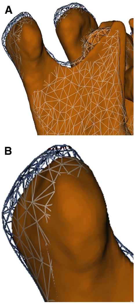
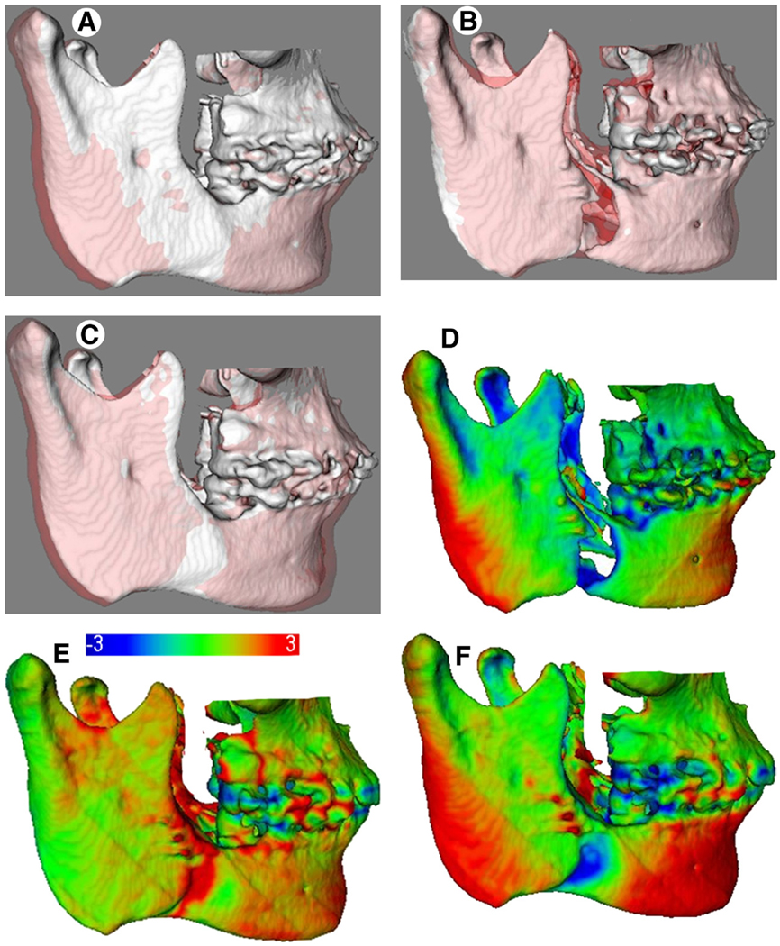
Patients and methods: This prospective observational study used preoperative and postoperative scans of 27 subjects presenting with a skeletal Class II jaw relationship with a normal or deep overbite. An automatic technique of cranial base superimposition was used to assess the positional and/or remodeling changes in the anatomic regions of interest. The displacements were visually displayed and quantified using 3-dimensional color maps. The positive and negative values of surface distances in the color maps indicated the direction of the displacements. Pearson correlation coefficients and a linear model for correlated data were used to evaluate the association between the regional displacements.
Results: The postoperative adaptations in the chin position between splint removal and 1 year after surgery were significantly negatively correlated with changes in the borders of the posterior ramus (left, r = -0.73, P ≤ .0001; and right, r = -0.68, P = .00) and the condyles (left, r = -0.53, P = .01; and right, r = -0.46, P = .02), indicating that these structures tended to be displaced in the same direction. Even though the mean condylar displacement with surgery was less than 1 mm, individual displacements greater than 2 mm with surgery were observed for 24% of the condyles. The condylar displacements were maintained at 1 year after surgery for 17% of the condyles.
Conclusions: The surface distance displacements indicated that the postoperative adaptations at different anatomic regions were significantly related.
Copyright © 2011 American Association of Oral and Maxillofacial Surgeons. Published by Elsevier Inc. All rights reserved.
Figures


References
-
- Proffit WR, Turvey TA, Phillips C. Orthognathic surgery: A hierarchy of stability. Int J Adult Orthodon Orthognath Surg. 1996;11:191. - PubMed
-
- Efstratiadis S, Baumrind S, Shofer F, et al. Evaluation of Class II treatment by cephalometric regional superimpositions versus conventional measurements. Am J Orthod Dentofac Orthop. 2005;128:607. - PubMed
-
- Costa F, Robiony M, Toro C, et al. Condylar positioning devices for orthognathic surgery: A literature review. Oral Surg Oral Med Oral Pathol Oral Radiol Endod. 2008;106:179. - PubMed
-
- Alder ME, Deahl ST, Matteson SR, et al. Short-term changes of condylar position after sagittal split osteotomy for mandibular advancement. Oral Surg Oral Med Oral Pathol Oral Radiol Endod. 1999;87:159. - PubMed
Publication types
MeSH terms
Grants and funding
LinkOut - more resources
Full Text Sources
