

Alcohol-related amnesia and dementia: animal models have revealed the contributions of different etiological factors on neuropathology, neurochemical dysfunction and cognitive impairment
- PMID: 21256970
- PMCID: PMC3086968
- DOI: 10.1016/j.nlm.2011.01.003
Alcohol-related amnesia and dementia: animal models have revealed the contributions of different etiological factors on neuropathology, neurochemical dysfunction and cognitive impairment
Abstract
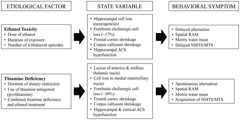
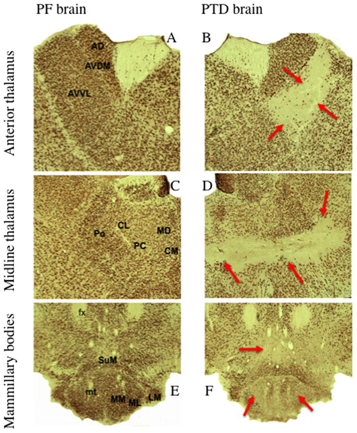
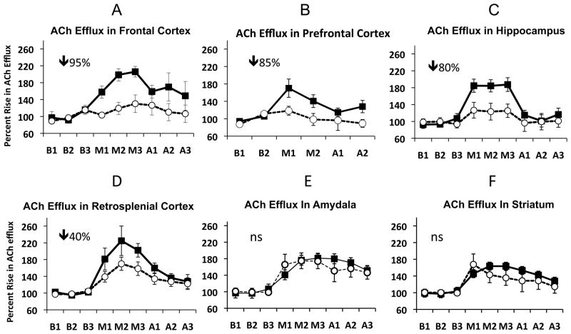
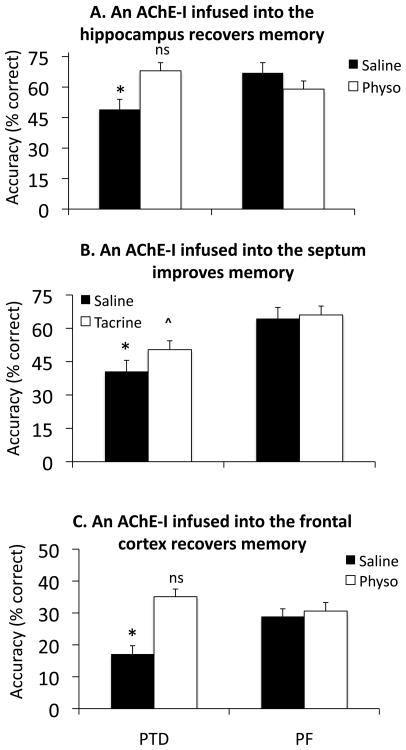
Chronic alcoholism is associated with impaired cognitive functioning. Over 75% of autopsied chronic alcoholics have significant brain damage and over 50% of detoxified alcoholics display some degree of learning and memory impairment. However, the relative contributions of different etiological factors to the development of alcohol-related neuropathology and cognitive impairment are questioned. One reason for this quandary is that both alcohol toxicity and thiamine deficiency result in brain damage and cognitive problems. Two alcohol-related neurological disorders, alcohol-associated dementia and Wernicke-Korsakoff syndrome have been modeled in rodents. These pre-clinical models have elucidated the relative contributions of ethanol toxicity and thiamine deficiency to the development of dementia and amnesia. What is observed in these models--from repeated and chronic ethanol exposure to thiamine deficiency--is a progression of both neural and cognitive dysregulation. Repeated binge exposure to ethanol leads to changes in neural plasticity by reducing GABAergic inhibition and facilitating glutamatergic excitation, long-term chronic ethanol exposure results in hippocampal and cortical cell loss as well as reduced hippocampal neurotrophin protein content critical for neural survival, and thiamine deficiency results in gross pathological lesions in the diencephalon, reduced neurotrophic protein levels, and neurotransmitters levels in the hippocampus and cortex. Behaviorally, after recovery from repeated or chronic ethanol exposure there is impairment in working or episodic memory that can recover with prolonged abstinence. In contrast, after thiamine deficiency there is severe and persistent spatial memory impairments and increased perseverative behavior. The interaction between ethanol and thiamine deficiency does not produce more behavioral or neural pathology, with the exception of reduction of white matter, than long-term thiamine deficiency alone.
Copyright © 2011 Elsevier Inc. All rights reserved.
Figures

Similar articles
-
Aging with alcohol-related brain damage: Critical brain circuits associated with cognitive dysfunction.Int Rev Neurobiol. 2019;148:101-168. doi: 10.1016/bs.irn.2019.09.002. Epub 2019 Oct 17. Int Rev Neurobiol. 2019. PMID: 31733663 Free PMC article. Review.
-
Interactions between chronic ethanol consumption and thiamine deficiency on neural plasticity, spatial memory, and cognitive flexibility.Alcohol Clin Exp Res. 2015 Nov;39(11):2143-53. doi: 10.1111/acer.12859. Epub 2015 Sep 30. Alcohol Clin Exp Res. 2015. PMID: 26419807 Free PMC article.
-
Aetiology of alcoholic brain damage: alcoholic neurotoxicity or thiamine malnutrition?Br Med Bull. 1994 Jan;50(1):99-114. doi: 10.1093/oxfordjournals.bmb.a072888. Br Med Bull. 1994. PMID: 8149203 Review.
-
Age-related vulnerability to diencephalic amnesia produced by thiamine deficiency: the role of time of insult.Behav Brain Res. 2004 Jan 5;148(1-2):93-105. doi: 10.1016/s0166-4328(03)00208-0. Behav Brain Res. 2004. PMID: 14684251
-
[Clinical application of neuroimaging to alcohol-related dementia].Nihon Arukoru Yakubutsu Igakkai Zasshi. 2012 Jun;47(3):125-34. Nihon Arukoru Yakubutsu Igakkai Zasshi. 2012. PMID: 22894053 Review. Japanese.
Cited by
-
Behavioral Neuroadaptation to Alcohol: From Glucocorticoids to Histone Acetylation.Front Psychiatry. 2016 Oct 6;7:165. doi: 10.3389/fpsyt.2016.00165. eCollection 2016. Front Psychiatry. 2016. PMID: 27766083 Free PMC article. Review.
-
Chronic voluntary alcohol consumption causes persistent cognitive deficits and cortical cell loss in a rodent model.Sci Rep. 2019 Dec 9;9(1):18651. doi: 10.1038/s41598-019-55095-w. Sci Rep. 2019. PMID: 31819151 Free PMC article.
-
PARP inhibition in vivo blocks alcohol-induced brain neurodegeneration and neuroinflammatory cytosolic phospholipase A2 elevations.Neurochem Int. 2019 Oct;129:104497. doi: 10.1016/j.neuint.2019.104497. Epub 2019 Jun 25. Neurochem Int. 2019. PMID: 31251945 Free PMC article.
-
The Neuroprotective Effects of Carvacrol on Ethanol-Induced Hippocampal Neurons Impairment via the Antioxidative and Antiapoptotic Pathways.Oxid Med Cell Longev. 2017;2017:4079425. doi: 10.1155/2017/4079425. Epub 2017 Jan 16. Oxid Med Cell Longev. 2017. PMID: 28191274 Free PMC article.
-
Dementia and osteoporosis in a geriatric population: Is there a common link?World J Orthop. 2017 May 18;8(5):412-423. doi: 10.5312/wjo.v8.i5.412. eCollection 2017 May 18. World J Orthop. 2017. PMID: 28567345 Free PMC article.
References
-
- Acker C. Neuropsychological deficits in alcoholics: the relative contributions of gender and drinking history. British Journal of Addiction. 1986;81:395–403. - PubMed
-
- Alexander-Kaufmann K, Harper C, Wilce P. Cerebellar vermis proteome of chronic alcoholic individuals. Alcoholism: Clinical and Experimental Research. 2007;31:1286–1296. - PubMed
-
- American Psychiatric Association. Diagnostic and statistical manual of mental disorders. 4. Washington, DC: American Psychiatric Association; 2000. pp. 199–205. Revised.
-
- Arendt T, Allen Y, Marchbanks RM, Schugens MM, Sinden J, Lantos PL, Gray JA. Cholinergic system and memory in the rat: Effects of chronic ethanol, embryonic basal forebrain brain transplants and excitotoxic lesions of cholinergic basal forebrain projection system. Neuroscience. 1989;33:435–462. - PubMed
Publication types
MeSH terms
Grants and funding
LinkOut - more resources
Full Text Sources
Medical
