. 2011 Apr;43(4):651-65.
doi: 10.1016/j.biocel.2011.01.008. Epub 2011 Jan 21.

c-Yes regulates cell adhesion at the blood-testis barrier and the apical ectoplasmic specialization in the seminiferous epithelium of rat testes

Affiliations

c-Yes regulates cell adhesion at the blood-testis barrier and the apical ectoplasmic specialization in the seminiferous epithelium of rat testes

Xiang Xiao et al. Int J Biochem Cell Biol. 2011 Apr.

Abstract

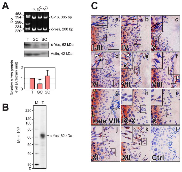
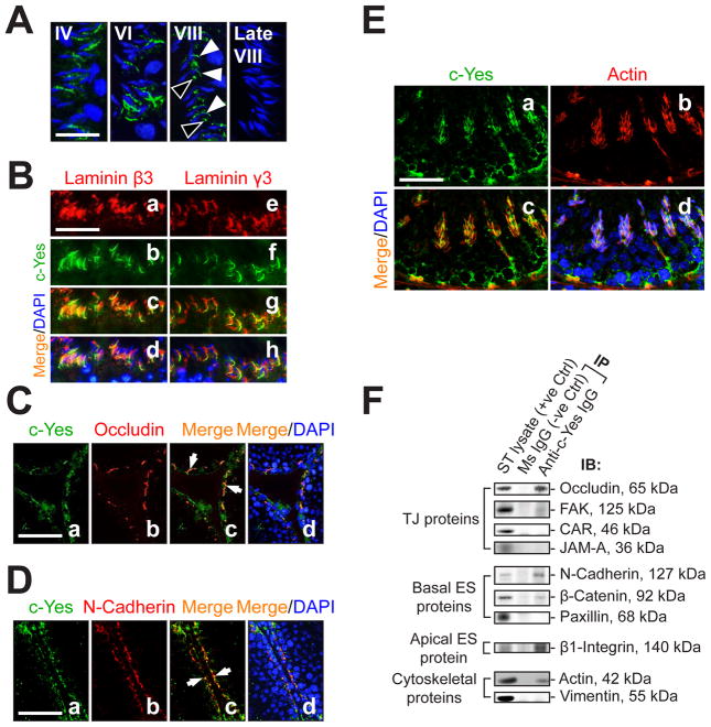
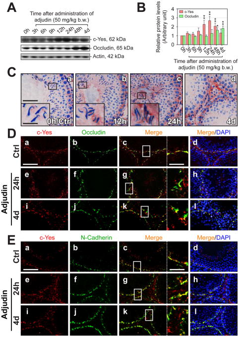
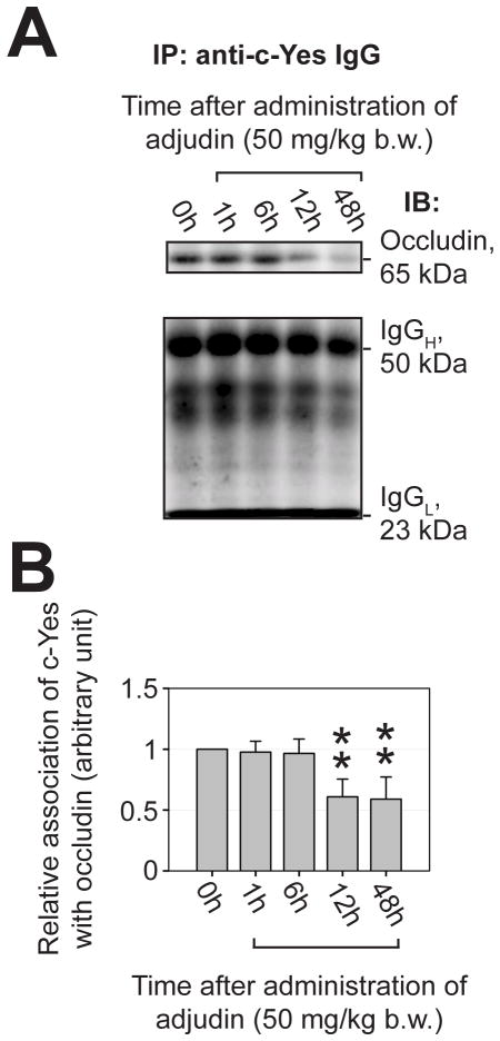
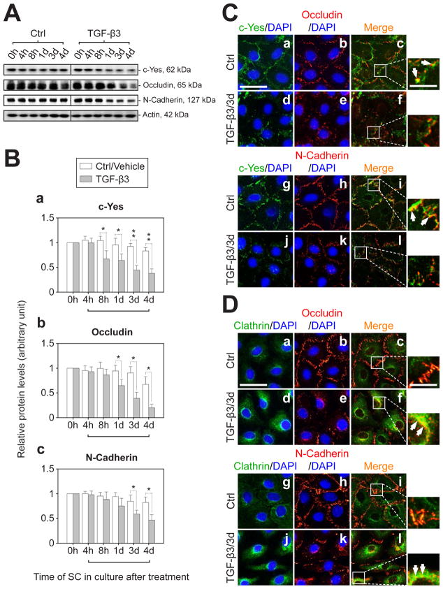
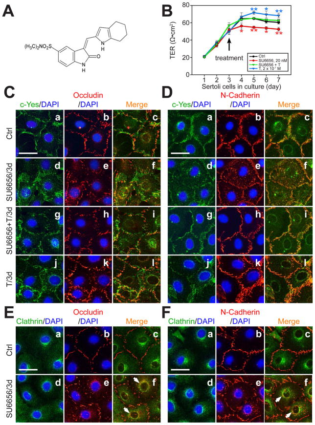
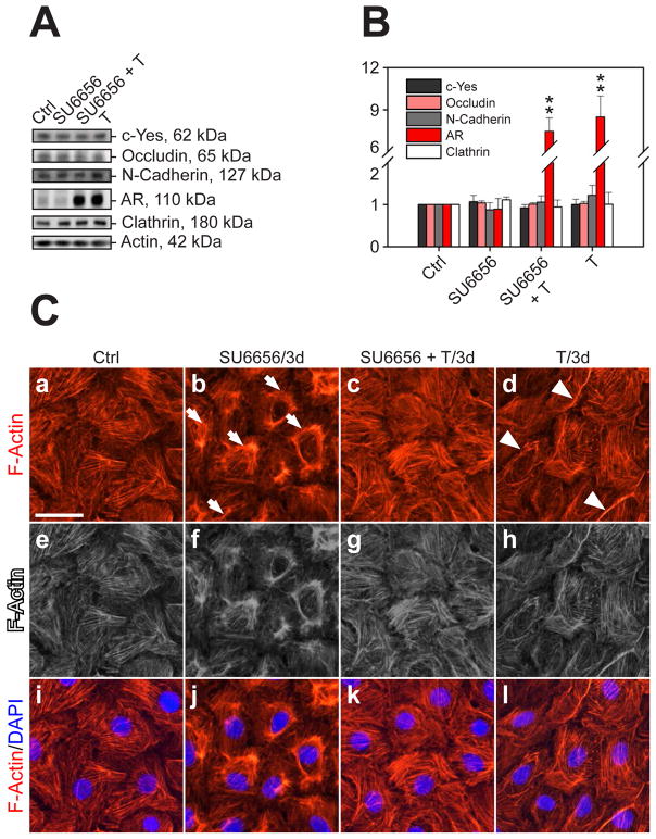
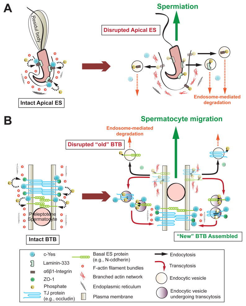
During spermatogenesis, extensive junction restructuring takes place at the blood-testis barrier (BTB) and the Sertoli cell-spermatid interface known as the apical ectoplasmic specialization (apical ES, a testis-specific adherens junction) in the seminiferous epithelium. However, the mechanism(s) that regulates these critical events in the testis remains unknown. Based on the current concept in the field, changes in the phosphorylation status of integral membrane proteins at these sites can induce alterations in protein endocytosis and recycling, causing junction restructuring. Herein, c-Yes, a non-receptor protein tyrosine kinase, was found to express abundantly at the BTB and apical ES stage-specifically, coinciding with junction restructuring events at these sites during the seminiferous epithelial cycle of spermatogenesis. c-Yes also structurally associated with adhesion proteins at the BTB (e.g., occludin and N-cadherin) and the apical ES (e.g., β1-integrin, laminins β3 and γ3), possibly to regulate phosphorylation status of proteins at these sites. SU6656, a selective c-Yes inhibitor, was shown to perturb the Sertoli cell tight junction-permeability barrier in vitro, which is mediated by changes in the distribution of occludin and N-cadherin at the cell-cell interface, moving from cell surface to cytosol, thereby destabilizing the tight junction-barrier. However, this disruptive effect of SU6656 on the barrier was blocked by testosterone. Furthermore, c-Yes is crucial to maintain the actin filament network in Sertoli cells since a blockade of c-Yes by SU6656 induced actin filament disorganization. In summary, c-Yes regulates BTB and apical ES integrity by maintaining proper distribution of integral membrane proteins and actin filament organization at these sites.


Figures

Fig. 1
Fig. 1. Cellular localization and stage-specific expression of c-Yes in the seminiferous epithelium of adult rat testes during the epithelial cycle of spermatogenesis
(A) Expression of c-Yes in testes (T) versus Sertoli (SC) and germ cells (GC) was assessed by RT-PCR (upper panel) and immunoblot analysis (middle panel) using either S16 or actin as a loading control. The relative c-Yes protein level in germ and Sertoli cells was compared with that in testes (50 μg protein per lane), of which the protein level was arbitrarily set at 1. Result is a mean ± SD of three independent experiments (lower panel). (B) An immunoblot demonstrating the specificity of an anti-c-Yes monoclonal antibody (mAb) (see Table 1) using lysate of rat testes (25 μg protein). Mr, Molecular weight; M, MagicMark XP Western Protein Standard (Invitrogen); D, dye front. (C) Frozen sections (7-μm thickness) of testes were used for immunohistochemistry (IHC) with an anti-c-Yes mAb shown in B, and representative tubules sorted by stages (I-XII) were shown in (a-k). The specificity of the c-Yes staining shown in (a-k) by IHC was validated and shown in (l) in which mouse IgG was used in place of the anti-c-Yes IgG. The expression of c-Yes is stage-specific, with the most intense staining in the epithelium near the basement membrane consistent with its localization at the BTB at stage VIII-IX (green arrowheads in g indicates the BTB site). c-Yes distribution in the seminiferous epithelium can also be discerned partly bordering the heads of elongating/elongated spermatids consistent with its localization at the apical ES except at late stage VIII when its expression diminished considerably (as magnified views from the boxed area in a-k), and c-Yes was found both at the convex (white arrowheads, d-f) and the concave (black arrowheads, d-e) sides at stages VI-VIII prior to spermiation. c-Yes was also found in the interstitial tissue (as the immune reaction toward the interstitium). c-Yes is a ubiquitously expressed tyrosine kinase, thus its staining pattern is diffused. Bar in a (also applies to b-l) = 55 μm; bar in insert in a (also applies to inserts in b-k) = 25 μm.
Fig. 2
Fig. 2. c-Yes is an integrated component of the BTB and the apical ES as demonstrated by dual- labeled immunofluorescence (IF) analysis and co-immunoprecipitation (Co-IP)
(A) Fluorescence microscopy was used to illustrate stage-specific localization of c-Yes (green, FITC) at the apical ES. Cell nuclei were visualized with DAPI (blue). c-Yes staining was detected at the apical ES surrounding the heads of the developing spermatids throughout the epithelial cycle until shortly before spermiation when it vanished completely (at late stage VIII). It is noted that c-Yes was found to be both at the convex (white arrowheads) and the concave (black arrowheads) sides in early stage VIII, consistent with IHC data shown in Fig. 1. Bar in first panel (also applies to the other three) = 30 μm. (B) Dual-labeled IF analysis confirmed c-Yes occurrence at the apical ES by its co-localization with laminin β3 (a-d) and γ3 (e-h) (red, Cy3), which are known apical ES components expressed exclusively by elongating/elongated spermatids (Yan and Cheng, 2006). Bar in a (also applies to b-h) = 60 μm. (C-D) Presence of c-Yes at the BTB was supported by moderate co-localization (white arrows) with TJ (occludin) (C) and basal ES (N-cadherin) (D) proteins. Bar in a (also applies to b-d) = 100 μm. (E) c-Yes presence at the BTB and the apical ES was further corroborated by its co-localization with F-actin, suggesting it is likely a regulatory component of the structural scaffolding of actin filaments. Bar in a (also applies to b-d) = 40 μm. (F) A study using Co-IP to assess the structural interactions between c-Yes and selected component proteins of the BTB and the apical ES using ~800 μg protein of seminiferous tubule (ST) lysates. Mouse (MS) IgG instead of anti-c-Yes IgG served as a negative control.
Fig. 3
Fig. 3. Changes in the steady-state level of c-Yes and its association with TJ and basal ES proteins at the BTB during adjudin-induced anchoring junction restructuring and germ cell depletion
(A) Testis lysates (50 μg protein) were used for immunoblotting to assess changes in the steady-state protein level of c-Yes, with occludin served as a positive control and actin a loading control. An induction of c-Yes expression was observed during the 12- to 48-h period after treatment. (B) A histogram with each bar shown as a mean ± SD of n = 3 rats using immunoblots such as those shown in (A). The protein levels of c-Yes and occludin at 0 hour were arbitrarily set at 1 and values were normalized and plotted against actin (*, P<0.05; **, P<0.01). (C) IHC staining of c-Yes obtained from the same rats after adjudin treatment used for immunoblot analysis. Within 12-h of treatment, some elongating/elongated spermatids were found to be misoriented and were depleting from the epithelium, even in a stage VII (C, b) tubule, and a considerable increase in c-Yes expression at the apical ES was detected in “prematurely” departing elongating spermatids in a stage VI (C, c) tubule (a-c, inset, magnified view of boxed area in the same panel). By 4 days following treatment, when virtually all tubules were devoid of elongating/elongated spermatids, c-Yes staining persisted at the BTB site. Bar in a (also applies to b-d) = 80 μm. Bar in inset in a = 20 μm, also applies insets in b and c. (D-E) IF staining showing c-Yes (red, Cy3) and BTB proteins occludin (green, FITC in D) and N-cadherin (green, FITC in E), and changes in their colocalization. Bar in a (also applies to b-l) = 120 μm. The staining patterns of c-Yes became “puffy” and less “compact” in the treatment groups versus control (normal) rats, and as a result, the association of c-Yes with these two BTB markers appeared to be considerably reduced, as shown in c, g, and k (inset at the right-hand partition with dotted lines is magnified view of boxed area in the same panel; bar = 40 μm). h, hour(s); d, day(s).
Fig. 4
Fig. 4. A loss of association between c-Yes and integral membrane protein occludin at the BTB after administration of adjudin to adult rats was examined by co-immunoprecipitation (Co-IP)
(A) c-Yes was first immunoprecipitated using an anti-c-Yes antibody with ~800 μg protein from corresponding tubule lysates obtained from rats treated with adjudin, and the changes in its interaction with occludin was quantified using Co-IP (see Materials and Methods) (upper panel). The lower panel shows the heavy (H) and the light (L) chains of IgG following Co-IP from the same blots shown on the upper panel to illustrate equal protein loading and uniform protein transfer. (B) Results of Co-IP experiments shown in (A) were summarized in this histogram. Each bar is a mean ± SD of three independent experiments. The relative association of c-Yes with occludin at 0 hour was arbitrarily set at 1 (*, P<0.05; **, P<0.01).
Fig. 5
Fig. 5. Changes in the steady-state protein level of c-Yes and its co-localization with BTB-associated proteins following treatment of Sertoli cell epithelium with TGF-β3
(A) Sertoli cells were cultured at a density of 0.5×10⁶ cells/cm2 for 4-d to allow the establishment of a functional TJ-barrier. Thereafter, the Sertoli cell epithelium was submitted to TGF-β3 treatment (3 ng/ml), with F12/DMEM replaced daily which also contained the desired amount of TGF-β3. Controls were Sertoli cells cultured with vehicle. Cell lysates were obtained for immunoblot analysis. Actin served as a protein loading control. (B) Results of the immunoblotting study, such as those shown in (A) were summarized in this histogram. The protein levels of c-Yes (a), occludin (b) and N-cadherin (c) at 0 h (hour) with or without TGF-β3 treatment were arbitrarily set at 1. Each bar is the mean ± SD of three independent experiments. Herein, the protein level after TGF-β3 treatment versus its corresponding control was compared (*, P<0.05; **, P<0.01). h, hour(s); d, day(s). (C) IF staining of Sertoli cells treated with TGF-β3 for 3-d to evaluate the cytokine effects on the distribution of c-Yes (green, FITC), occludin (red, Cy3, a-f) and N-cadherin (red, Cy3, g-l) and the changes in their co-localization pattern. Besides a TGF-β3-induced decline in the levels of c-Yes, occludin and N-cadherin, treatment of cells with TGF-β3 also resulted in a loss of interactions between c-Yes and the integral membrane proteins at the BTB, because of the result of protein internalization, as indicated in smaller panels on the right-hand column, which are magnified views of the boxed area in c, f, i, and l (see f vs. c and l vs. i). White arrows illustrate the explicit co-localization of corresponding proteins in the same row. (D) Since the BTB marker proteins occludin and N-cadherin appeared to become internalized following TGF-β3 treatment, an endocytic vesicle marker clathrin (green, FITC) was used in this dual-labeled IF analysis to examine if the changes noted in C was mediated via endocytosis, and indeed occludin (red, Cy3, a-f) and N-cadherin (red, Cy3, g-l) were found to localize to this endocytic component more extensive after TGF-β3 treatment (see white arrows in the right panels in D). Bar in a (also applies to b-l in C-D) = 55 μm, wherein bar = 20 μm for the magnified box area in c, f, i and l in C and D, which applies to all micrographs in this column on the right panel.
Fig. 6
Fig. 6. A study to assess the effects of SU6656 on the Sertoli cell TJ-permeability barrier and protein distribution at the Sertoli-Sertoli cell interface
(A) Structural formula of SU6656. (B) Sertoli cells were cultured at a density of 2×10⁶ cells/cm2 on Matrigel-coated bicameral units and the TJ barrier function was monitored by quantifying the TER across the Sertoli cell epithelium. On day 3, Sertoli cells in the bicameral units were treated with either SU6656 (2×10⁻⁸ M), testosterone (T, 2×10⁻⁷ M), or SU6656 + T (see arrow). After TER reading, media were replaced with fresh F12/DMEM containing the desired amount of either SU6656 or T. Each data point is a mean ± SD of quadruplicates of a representative experiment, and this experiment was repeated three times using different batches of Sertoli cells and yielded similar results (*, P<0.05; **, P<0.01). (C, D) Dual-labeled IF analysis to assess changes on the protein distribution of either occludin (C) or N-cadherin (D) at the cell-cell interface using Sertoli cells cultured at 0.05 × 10⁶ cells/cm2 on Matrigel-coated coverslips and cells were subjected to the same treatments as shown in B. Bar in a (also applies to b-l) = 45 μm. (E, F) Dual-labeled IF analysis was used to confirm the internalization of BTB proteins occludin and N-cadherin by their increase in partial co-localization with clathrin after SU6656 treatment. Bar in a (applies to b-f) = 45 μm. White arrows illustrate co-localization of either occludin or N-cadherin with clathrin.
Fig. 7
Fig. 7. A study to assess the effects of SU6656 on the steady-state protein levels of c-Yes and other BTB-associated proteins and cellular redistribution of F-actin in the Sertoli cell epithelium
(A) Immunoblot analysis to assess changes in the protein levels of c-Yes, occludin, N-cadherin, androgen receptor (AR) and clathrin after treatment of SU6656, T or SU6656 + T. (B) Histogram of the immunoblot analysis from three independent experiments normalized against actin (**, P<0.01). (C) Sertoli cells were cultured at 0.025–0.04 × 10⁶ cells/cm2 on Matrigel-coated coverslips for 1.5~2 days, forming an intact epithelium with ultrastructures of TJ and basal ES (Siu et al., 2005), before treatment. After 3-day incubation with SU6656, T or SU6656 + T, cells were stained with rhodamine phalloidin to visualize F-actin (red, Cy3). In comparison with controls, F-actin filaments in cells treated with SU6656 became considerably disorganized, and withdrew from the cell cytosol and more concentrated toward the nuclei (arrows in b) (see b and j vs. a and i, blue, DPAI). Gray scale images shown in e-h were used to better illustrate the changes shown in a-d vs. i-l. However, the presence of T was shown to block the disruptive effects of SU6656 on the actin filament network in Sertoli cells when compared to control cells (see c and k vs. b and j, a and i). In cells treated with T alone, the F-actin filament network was similar to control cells but the network appeared to be fortified at the cell-cell interface (see white arrowheads in d), apparently to promote the Sertoli cell TJ-permeability barrier function as shown in Fig. 6.
Fig. 8
Fig. 8. A schematic drawing illustrating the underlying mechanism by which c-Yes regulates apical ES and BTB restructuring during the seminiferous epithelial cycle of spermatogenesis
On the left panels of (A) and (B) are the corresponding intact apical ES and BTB (e.g., stage VII) in the epithelium of adult rat testes, with c-Yes highly expressed and tightly associated with the corresponding cell adhesion protein complexes at these sites, such as apical ES proteins: β1-integin; and BTB proteins: occludin and N-cadherin. When apical ES undergoes disruption to prepare for spermiation at stage VIII of the epithelial cycle (right panel in A), c-Yes likely induces protein phosphorylation, causing protein endocytosis to destabilize the apical ES to facilitate spermiation. Similar events take place at the “old” BTB site above the preleptotene spermatocytes in transit in which c-Yes induces phosphorylation of occludin and N-cadherin, which in turn become internalized via endocytic vesicle-mediated pathway likely mediated by cytokines (e.g., TGF-β2, TNFα), thereby destabilizing the “old” BTB site to facilitate the transit of spermatocytes. However, c-Yes is possibly working in concert with androgen which is known to promote BTB integrity (such as via de novo synthesis of “new” TJ proteins to be assembled to the “new” TJ-fibrils below the preleptotene spermatocytes in transit to assemble a “new” BTB) to establish a “new” BTB below the spermatocytes (right panel in B, see also text for details). Additionally, other proteins (e.g., 14-3-3) (Wong et al., 2009) and testosterone (Yan et al., 2008) promote protein transcytosis, most likely from the “old” to the “new” BTB site (see right panel in B). In short, this provides a unique mechanism to maintain the immunological barrier and seminiferous epithelial integrity during the epithelial cycle of spermatogenesis.

Similar articles

Cited by

References

    1. Blake RA, Broome MA, Liu X, Wu J, Gishizky M, Sun L, et al. SU6656, a selective src family kinase inhibitor, used to probe growth factor signaling. Mol Cell Biol. 2000;20:9018–27. - PMC - PubMed
    1. Boutros T, Chevet E, Metrakos P. Mitogen-activated protein (MAP) kinase/MAP kinase phosphatase regulation: Roles in cell growth, death, and cancer. Pharmacol Rev. 2008;60:261–310. - PubMed
    1. Bowman T, Broome MA, Sinibaldi D, Wharton W, Pledger WJ, Sedivy JM, et al. Stat3-mediated Myc expression is required for Src transformation and PDFG-induced mitogenesis. Proc Natl Acad Sci USA. 2001;98:7319–24. - PMC - PubMed
    1. Chen YH, Lu Q, Goodenough DA, Jeansonne B. Nonreceptor tyrosine kinase c-Yes interacts with occludin during tight junction formation in canine kidney epithelial cells. Mol Biol Cell. 2002;13:1227–37. - PMC - PubMed
    1. Cheng CY, Mruk DD. Cell junction dynamics in the testis: Sertoli-germ cell interactions and male contraceptive development. Physiol Rev. 2002;82:825–74. - PubMed

Publication types

MeSH terms

LinkOut - more resources