

Bicuspid aortic valve disease: the role of oxidative stress in Lrp5 bone formation
- PMID: 21257323
- PMCID: PMC3085628
- DOI: 10.1016/j.carpath.2010.11.007
Bicuspid aortic valve disease: the role of oxidative stress in Lrp5 bone formation
Abstract
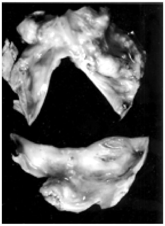
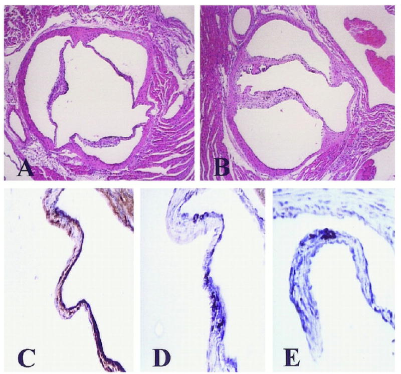
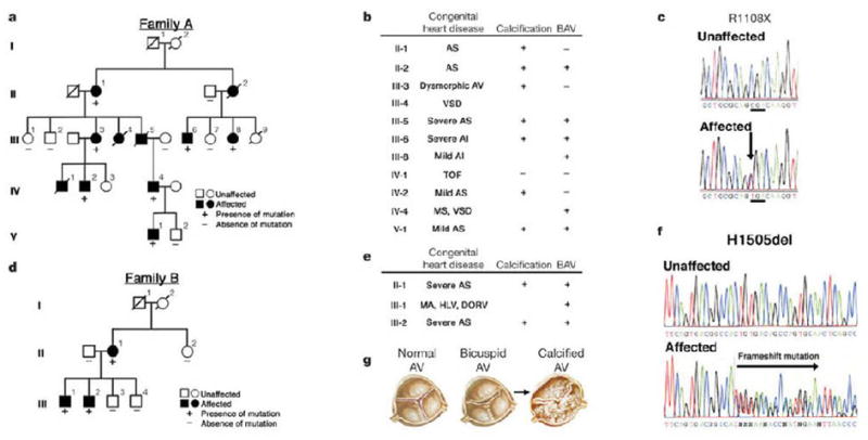
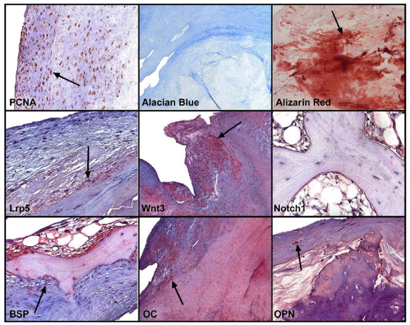
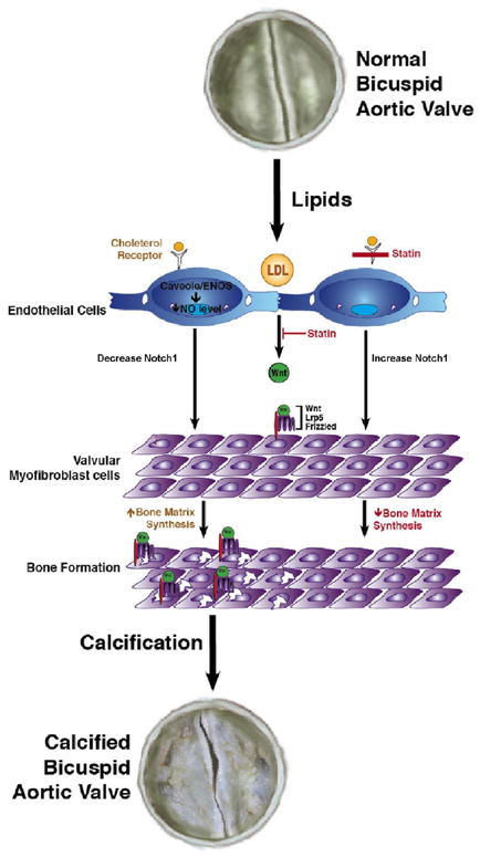
The bicuspid aortic valve is a common congenital cardiac anomaly, having a prevalence of 0.9% to 1.37% in the general population and a male preponderance ratio of 2:1. The recognition of a bicuspid aortic valve is clinically relevant because of its association with aortic stenosis or regurgitation, aortic aneurysm or dissection, and infective endocarditis. Although some patients with a bicuspid aortic valve may go undetected without clinical complications for a lifetime, the vast majority will require intervention, most often surgery, at some point. In fact, the natural history of bicuspid aortic valve is that of valve calcification and progressive stenosis that typically occur at a faster rate than in tricuspid aortic valves. This pattern of presentation supports the hypothesis that shear stress in patients with congenitally abnormal aortic valves may contribute to an earlier leaflet calcification. However, there is emerging research data showing that the valve calcification process might have a similar pathophysiologic process to that of vascular atherosclerosis. This review focuses on the current knowledge of the cellular mechanisms of bicuspid aortic valve.
Copyright © 2011 Elsevier Inc. All rights reserved.
Figures
References
-
- Rajamannan NM, Subramaniam M, Springett M, Sebo TC, Niekrasz M, McConnell JP, Singh RJ, Stone NJ, Bonow RO, Spelsberg TC. Atorvastatin inhibits hypercholesterolemia-induced cellular proliferation and bone matrix production in the rabbit aortic valve. Circulation. 2002;105(22):2260–2265. - PMC - PubMed
-
- Rajamannan NM, Nealis TB, Subramaniam M, Pandya S, Stock SR, Ignatiev CI, Sebo TJ, Rosengart TK, Edwards WD, McCarthy PM, Bonow RO, Spelsberg TC. Calcified rheumatic valve neoangiogenesis is associated with vascular endothelial growth factor expression and osteoblast-like bone formation. Circulation. 2005;111(24):3296–3301. - PMC - PubMed
-
- Roberts WC. The congenitally bicuspid aortic valve. A study of 85 autopsy cases. The American Journal of Cardiology. 1970;26:72–83. - PubMed
-
- Roberts WC, Ko JM. Frequency by decades of unicuspid, bicuspid, and tricuspid aortic valves in adults having isolated aortic valve replacement for aortic stenosis, with or without associated aortic regurgitation. Circulation. 2005;111(7):920–925. - PubMed
Publication types
MeSH terms
Substances
Grants and funding
LinkOut - more resources
Full Text Sources
Other Literature Sources
Medical
