. 2011 May;25(5):1556-66.
doi: 10.1096/fj.10-177279. Epub 2011 Jan 21.

Proinflammatory role of aquaporin-4 in autoimmune neuroinflammation

Affiliations

Proinflammatory role of aquaporin-4 in autoimmune neuroinflammation

Lihua Li et al. FASEB J. 2011 May.

Abstract

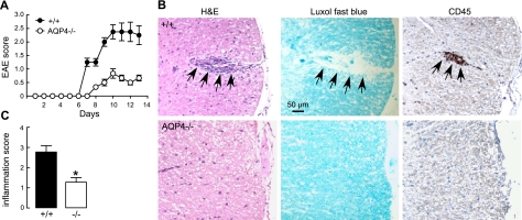
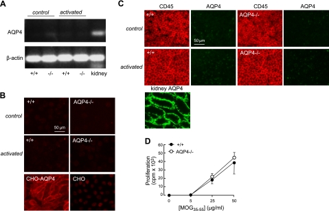
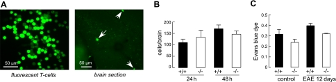
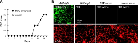
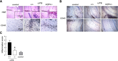
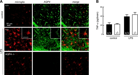
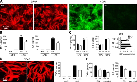
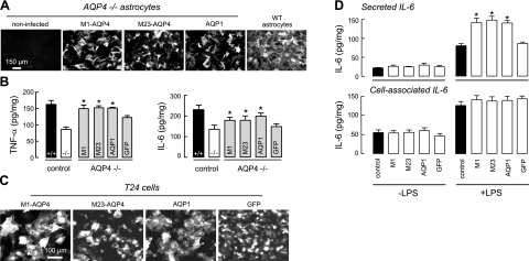
Aquaporin-4 (AQP4) deficiency in mice reduces neuroinflammation in experimental autoimmune encephalomyelitis (EAE) produced by active immunization with myelin oligodendrocyte glycoprotein peptide (MOG). Potential mechanisms for the protective effect of AQP4 deficiency were investigated, including AQP4-dependent leukocyte and microglia cell function, immune cell entry in the central nervous system (CNS), intrinsic neuroinflammation, and humoral immune response. As we found with active-immunization EAE, neuroinflammation was greatly reduced in AQP4-knockout mice in adoptive-transfer EAE. AQP4 was absent in immune cells, including activated T lymphocytes. The CNS migration of fluorescently labeled, MOG-sensitized T lymphocytes was comparable in wild-type and AQP4-knockout mice. Microglia did not express AQP4. Serum anti-AQP4 antibodies were absent in EAE. Remarkably, intracerebral injection of LPS produced much greater neuroinflammation in wild-type than in AQP4-knockout mice, and cytokine (TNF-α and IL-6) secretion was reduced in astrocyte cultures from AQP4-knockout mice. Adenovirus-mediated expression of AQP4, or of an unrelated aquaporin, AQP1, increased cytokine secretion in astrocyte and nonastrocyte cell cultures, supporting the involvement of aquaporin water permeability in cytokine secretion. Our data suggest an intrinsic proinflammatory role of AQP4 involving AQP4-dependent astrocyte swelling and cytokine release. Reduction in AQP4 water transport may be protective in neuroinflammatory CNS diseases.


Figures

Figure 1.
Figure 1.
Attenuated adoptive-transfer EAE in AQP4-null mice. A) Activated, MOG-sensitized T lymphocytes from wild-type mice were transferred to naive wild-type (+/+) or AQP4-null (−/−) mice. Means ± se of clinical scores (4 mice/group). B) Representative spinal cord H&E staining (left panels), Luxol fast blue staining (middle panels), and CD45 immunocytochemistry (right panels) at 15 d after T-lymphocyte transfer into wild-type or AQP4-null mice. Arrows denote EAE lesion. C) Inflammatory score determined by masked assessment of H&E and CD45 sections (se, 3 mice/group, 5 sections examined/mouse). *P < 0.001.
Figure 2.
Figure 2.
Absence of AQP4 expression in immune cells. A) RNA was isolated from T lymphocytes, without (control) or after MOG sensitization (as done for adoptive-transfer EAE), and from mouse kidney (positive control). AQP4 and β-actin were amplified by RT-PCR using specific primers. Representative of 3 sets of amplifications on different cultures. B) AQP4 immunofluorescence in T lymphocytes, without or after MOG sensitization. AQP4-expressing Chinese hamster ovary (CHO) cells (CHO-AQP4) as the positive control and nontransfected CHO cells (CHO) as the negative control. C) CD45 and AQP4 immunofluorescence in lymph nodes of control mice and at 15 d after MOG immunization. Mouse kidney shown as a positive control for AQP4. D) Splenocytes from wild-type and AQP4-deficient mice stimulate proliferation of MOG35–55-specific T lymphocyte in a similar manner. MACS-separated 2D2 T lymphocytes were cultured with irradiated splenic antigen-presenting cells from naive wild-type or AQP4-deficient mice in the presence of MOG35–55. Proliferation was measured by [3H]-thymidine incorporation (sd, 3 sets of cultures, differences not significant).
Figure 3.
Figure 3.
AQP4 deletion does not alter penetration of T lymphocytes into the CNS. MOG-sensitized T lymphocytes (as prepared for adoptive-transfer EAE) were fluorescently labeled with CFSE. Naive wild-type and AQP4-null mice were injected intravenously with 2 × 10⁷ labeled lymphocytes, and brains were perfused and harvested at 24 and 48 h. A) Left panel: fluorescently labeled MOG-activated T lymphocytes. Right panel: representative frozen section of brain, showing fluorescent T lymphocytes that crossed the blood-brain barrier. B) Number of fluorescent T lymphocytes counted in 10-μm-thick brain sections per mouse at 24 and 48 h (se, 3 mice/group at each time point, differences not significant). C) Extravasated Evans blue dye in brain in control mice and at 12 d after MOG immunization (se, 4 mice/group at each time point, differences not significant).
Figure 4.
Figure 4.
Absence of anti-AQP4 autoantibodies in EAE. A) Active-immunization EAE was produced in wild-type mice by MOG35–55 peptide. Clinical score. B) AQP4-expressing FRT cells (FRT-AQP4) and control nontransfected cells (FRT) stained with serum from control and EAE mice, and green fluorescent secondary anti-mouse antibody. Positive control is human serum from patient with NMO (EAE serum). AQP4 stained red. Representative of 3 sets of staining studies.
Figure 5.
Figure 5.
Reduced brain inflammation in AQP4-null mice after intracerebral LPS injection. Mice were injected intracerebrally with PBS (control) or LPS. A) Brain histology at 1 d by H&E staining (top panels) and CD45 immunocytochemistry (bottom panels). B) Gallery of CD45-immunostained sections of hippocampus from 3 wild-type and 3 AQP4-null mice. C) Quantification of inflammation done using H&E and CD45-stained sections (se). *P < 0.01 vs. +/+.
Figure 6.
Figure 6.
Microglial cells do not express AQP4. A) Absence of AQP4 expression in reactive microglia following intracerebral injection of LPS. Immunofluorescence of brain sections stained for microglial marker anti-Iba1 (red) and AQP4 (green). Representative of 3 sets of experiments. Scale bars = 50 μm. B) TNF-α in whole-brain homogenates at 2 h after intracerebral injection of saline (control) or LPS (se, n=4, differences between +/+ and −/− not significant).
Figure 7.
Figure 7.
Reduced cytokine release by astrocyte cultures in AQP4 deficiency. A) GFAP (left panels) and AQP4 (right panels) immunofluorescence of differentiated, primary cultures of astrocytes from neonatal mouse brain cortex. B) TNF-α and IL-6 in culture medium at 2 h after LPS (100 ng/ml) or saline addition (se, n=6). Representative of 5 sets of cultures. C) Cell-associated TNF-α and IL-6 at 2 h after LPS (se, n=4, differences not significant comparing −LPS and +LPS). Representative of 3 sets of cultures. (right) Quantitative real-time RT-PCR of indicated transcripts from astrocyte cultures (se, n=4, differences not significant comparing −LPS and +LPS). D) Right panel: TNF-α in culture medium at 24 h after Mn3+ exposure (se, n=4). Left panels: GFAP immunofluorescence of astrocytes after 24 h Mn3+ exposure. E) TNF-α and IL-6 in culture medium at 2 h after LPS in control medium (290 mosmol) or hyperosmolar (310 mosmol) medium containing excess 20 mM mannitol (se, n=4). *P < 0.01 vs. corresponding +/+.
Figure 8.
Figure 8.
Evidence for aquaporin-facilitated cytokine secretion. A) Immunofluorescence of AQP4 (M1 and M23 isoforms) and AQP1 in adenovirus-treated primary astrocyte cultures from AQP4-null mice (AQP4−/− astrocytes). AQP4 immunofluorescence of culture from wild-type mice shown at right. B) TNF-α and IL-6 in culture medium at 2 h after LPS (100 ng/ml) or saline addition (se, n=4). Representative of 3 sets of cultures/infections. *P < 0.01 vs. −/− control. C) AQP4 and AQP1 immunofluorescence, and GFP fluorescence, of adenovirus-treated T24 (bladder) cells. D) Secreted IL-6 (in culture medium, top panel) and cell-associated IL-6 (bottom panel) at 2 h after addition of LPS (se, n=4). *P < 0.01 vs. LPS-treated control.

Similar articles

Cited by

References

    1. Ransohoff R. M. (2006) A mighty mouse: building a better model of multiple sclerosis. J. Clin. Invest. 116, 2313–2316 - PMC - PubMed
    1. Korn T., Bettelli E., Oukka M., Kuchroo V. K. (2009) IL-17 and Th17 cells. Annu. Rev. Immunol. 27, 485–517 - PubMed
    1. Kroenke M. A., Carlson T. J., Andjelkovic A. V., Segal B. M. (2008) IL-12- and IL-23-modulated T cells induce distinct types of EAE based on histology, CNS chemokine profile, and response to cytokine inhibition. J. Exp. Med. 205, 1535–1541 - PMC - PubMed
    1. Lyons J. A., Ramsbottom M. J., Cross A. H. (2002) Critical role of antigen-specific antibody in experimental autoimmune encephalomyelitis induced by recombinant myelin oligodendrocyte glycoprotein. Eur. J. Immunol. 32, 1905–1913 - PubMed
    1. Weber M. S., Prod'homme T., Patarroyo J. C., Molnarfi N., Karnezis T., Lehmann-Horn K., Danilenko D. M., Eastham-Anderson J., Slavin A. J., Linington C., Bernard C. C., Martin F., Zamvil S. S. (2010) B-cell activation influences T-cell polarization and outcome of anti-CD20 B-cell depletion in central nervous system autoimmunity. Ann. Neurol. 68, 369–383 - PMC - PubMed

Publication types

MeSH terms

LinkOut - more resources