. 2011 Apr;25(4):1284-94.
doi: 10.1096/fj.10-160051. Epub 2011 Jan 21.

Noninvasive molecular imaging reveals role of PAF in leukocyte-endothelial interaction in LPS-induced ocular vascular injury

Affiliations

Noninvasive molecular imaging reveals role of PAF in leukocyte-endothelial interaction in LPS-induced ocular vascular injury

Rebecca C Garland et al. FASEB J. 2011 Apr.

Abstract

Uveitis is a systemic immune disease and a common cause of blindness. The eye is an ideal organ for light-based imaging of molecular events underlying vascular and immune diseases. The phospholipid platelet-activating factor (PAF) is an important mediator of inflammation, the action of which in endothelial and immune cells in vivo is not well understood. The purpose of this study was to investigate the role of PAF in endothelial injury in uveitis. Here, we use our recently introduced in vivo molecular imaging approach in combination with the PAF inhibitors WEB 2086 (WEB) and ginkgolide B (GB). The differential inhibitory effects of WEB and GB in reducing LPS-induced endothelial injury in the choroid indicate an important role for PAF-like lipids, which might not require the PAF receptor for their signaling. P-selectin glycoprotein ligand-1-mediated rolling of mouse leukocytes on immobilized P-selectin in our autoperfused microflow chamber assay revealed a significant reduction in rolling velocity on the cells' contact with PAF. Rolling cells that came in contact with PAF rapidly assumed morphological signs of cell activation, indicating that activation during rolling does not require integrins. Our results show a key role for PAF in mediating endothelial and leukocyte activation in acute ocular inflammation. Our in vivo molecular imaging provides a detailed view of cellular and molecular events in the complex physiological setting.


Figures

Figure 1.
Figure 1.
PAF-induced leukocyte adhesion in retinal and choroidal vessels. PAF (5 μg) was injected into the vitreous of Lewis rats; 2 h later, animals were perfused, and the vasculature was ConA stained. Subsequently, retinal and choroidal flat mounts were prepared, and number of firmly adhering leukocytes was quantified by counting. A) Representative micrographs of firmly adhering leukocytes (arrows) in retinal vessels. Higher magnification micrographs (bottom panels) show sections of the retinal veins. B) Numbers of leukocytes in retinal arteries. C) Numbers of leukocytes in retinal veins. D) Numbers of firmly adhering leukocytes in choroidal vessels. Bars are means ± se.
Figure 2.
Figure 2.
Effect of PAF inhibition on leukocyte accumulation in retinal and choroidal vessels of EIU rats. To inhibit PAF, animals were intraperitoneally treated with WEB (2.5 mg), GB (2.5 mg), or both; 30 min minutes later, EIU was induced by toepad injection of LPS (100 μg/kg), and 24 h later, animals were perfused with rhodamine-conjugated ConA to remove the intravascular content and stain vessels and firmly adhering leukocytes. A) Representative micrographs of adherent leukocytes (arrows) in retinas from control and PAF-inhibitor-treated EIU rats. Higher magnification micrographs (bottom panels) show sections of the retinal veins. B) Quantification of firmly adhering leukocytes in retinal arteries. C) Firmly adhering leukocytes in retinal veins. D) Firmly adhering leukocytes in choroidal vessels.
Figure 3.
Figure 3.
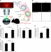
Molecular imaging with rPSGL-1-conjugated fluorescent MSs reveals role of PAF in endothelial injury in vivo. Endothelial injury in the fundus vasculature in vivo was quantified using our adhesion-molecule-conjugated imaging agents in normal animals with and without intravitreal PAF injection. At 24 h after PAF-injection, rPSGL-1-conjugated MSs were systemically injected; 30 min later, the fundus was imaged using SLO (30° angle at 15 frames/s). Number of MSs in this region was quantified. A) Schematic of our in vivo molecular imaging approach. B) Representative SLO still images showing adhering MSs in retinas of live animals. Bright spots indicate adhering MSs that resisted the blood flow. C) In vivo quantification of the number of accumulated MSs in retinal vessels, indicating the level of endothelial injury. D) In vivo quantification of MS accumulation in the choroidal vessels of control and PAF-injected animals. E) Ex vivo quantification of the number of MSs in flat mounts of retinal vessels. F) Ex vivo quantification of the number of MSs in flat mounts of choroidal vessels.
Figure 4.
Figure 4.
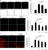
Effect of PAF inhibition on retinal and choroidal P-selectin up-regulation. To quantify the level of P-selectin expression in retinal and choroidal endothelium of normal and EIU rats, with and without PAF inhibition, we injected rPSGL-1-conjugated MSs, 24 h after LPS injection, and performed in vivo molecular imaging of the fundus vessels or ex vivo epifluorescence microscopy of retinal and choroidal flat mounts. A) In vivo binding analysis in choroidal vessels was performed 30 min after MS injection. B) Quantification of MS binding to choroidal microvessels in normal and EIU rats with or without inhibitor pretreatment. C) Representative retinal flat mounts from normal and EIU animals with or without inhibitor treatments. Bright green spots, adhering MSs; round red cells in vessels, firmly adhering leukocytes. D) Quantification of the number of accumulated rPSGL-1-conjugated MSs in the retinal vessels of normal and EIU animals with or without PAF inhibitor treatment. E) Representative choroidal flat mounts from normal and EIU animals with or without inhibitor treatments. F) Quantification of the number of accumulated rPSGL-1-conjugated MSs in the choroidal vessels of normal and EIU animals with or without PAF inhibitor treatment. Bars are means ± se.
Figure 5.
Figure 5.
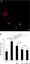
Ratios of number of MSs binding to leukocytes to that of total MSs counted. To distinguish the binding of rPSGL-1-conjugated MSs to activated endothelial cells, from those bound to adherent leukocytes, we calculated the ratio between them under various conditions. A) Confocal micrograph showing adhesion of an MS to a firmly adhering leukocyte. B) Ratios of MSleu to MStotal showing the effect of EIU and PAF inhibitors on MS binding to leukocytes.
Figure 6.
Figure 6.
Effect of PAF inhibitors on leukocyte and MS binding to retinal and choroidal vasculatures of normal rats. To investigate the constitutive role of PAF in endothelial function, the level of endothelial activation and leukocyte adhesion was quantified in animals 24 h after treatments. Adherent leukocytes were counted in retinal and choroidal flat mounts 24 h after intraperitoneal injection of PAF inhibitors to normal rats. A) Number of firmly adhering leukocytes in retinal arteries after GB intraperitoneal injection. B) Number of firmly adhering leukocytes in retinal veins after GB intraperitoneal injection. C) Number of bound rPSGL-1-conjugated MSs in retinal vessels after injection of WEB or GB. D) Number of bound MSs in the choroid of control animals and in those pretreated with WEB or GB.
Figure 7.
Figure 7.
Direct effect of PAF on PSGL-1-mediated rolling in vivo. Velocity of the rolling leukocytes was analyzed in vivo as they passed through the transparent P-selectin-coated, chamber. Blood is pumped by the mouse heart through a biocompatible tube from the carotid artery to the chamber and then to the jugular vein on the opposite side. Ports in the tube provide a site for blood pressure monitoring and control as well as one for injecting experimental agents. A) Schematic of the experimental design illustrating the study of the immediate effect of PAF on rolling leukocytes, originally introduced by Hafezi-Moghadam et al. (17). B) Successive in vivo images of a rolling leukocyte on immobilized P-selectin before (top panel) and one after PAF infusion (0.6 μg in 100 μl saline; bottom panel). C) Cumulative histogram of the velocity of rolling leukocytes on immobilized P-selectin without, and 10 min after, PAF infusion. D) Representative micrographs of activated leukocytes (arrows) bound to P-selectin 10 min after PBS control or PAF infusion.

References

    1. Suzuma K., Mandai M., Kogishi J., Tojo S. J., Honda Y., Yoshimura N. (1997) Role of P-selectin in endotoxin-induced uveitis. Invest. Ophthalmol. Vis. Sci. 38, 1610–1618 - PubMed
    1. Miyamoto K., Ogura Y., Hamada M., Nishiwaki H., Hiroshiba N., Honda Y. (1996) In vivo quantification of leukocyte behavior in the retina during endotoxin-induced uveitis. Invest. Ophthalmol. Vis. Sci. 37, 2708–2715 - PubMed
    1. Devine L., Lightman S. L., Greenwood J. (1996) Role of LFA-1, ICAM-1, VLA-4 and VCAM-1 in lymphocyte migration across retinal pigment epithelial monolayers in vitro. Immunology 88, 456–462 - PMC - PubMed
    1. Norman K. E., Moore K. L., McEver R. P., Ley K. (1995) Leukocyte rolling in vivo is mediated by P-selectin glycoprotein ligand-1. Blood 86, 4417–4421 - PubMed
    1. Moore K. L., Patel K. D., Bruehl R. E., Li F., Johnson D. A., Lichenstein H. S., Cummings R. D., Bainton D. F., McEver R. P. (1995) P-selectin glycoprotein ligand-1 mediates rolling of human neutrophils on P-selectin. J. Cell Biol. 128, 661–671 - PMC - PubMed

Publication types

LinkOut - more resources