. 2011;60(1):89-93.
doi: 10.1093/jmicro/dfq081. Epub 2011 Jan 21.

Direct imaging of pH1N1 2009 influenza virus replication in alveolar pneumocytes in fatal cases by transmission electron microscopy

Affiliations

Direct imaging of pH1N1 2009 influenza virus replication in alveolar pneumocytes in fatal cases by transmission electron microscopy

Atanu Basu et al. J Electron Microsc (Tokyo). 2011.

Abstract

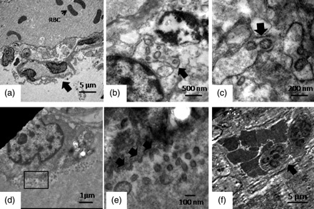
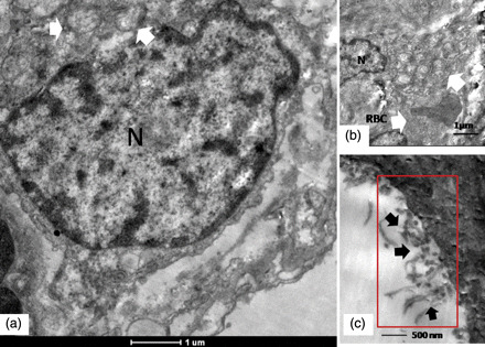
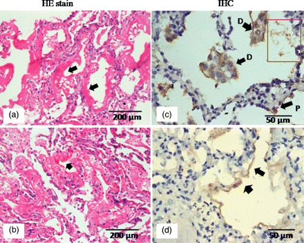
Human influenza virus pandemics constitute a major global public health issue. Although studies on autopsy specimens from the recent pandemic by the 2009 influenza A (H1N1) virus have revealed a broad spectrum of pathologic findings, direct electron microscopic studies of the lung tissue from influenza fatalities are few. In this study, we examined five well-preserved pulmonary necropsy specimens from fatal cases of laboratory-confirmed pH1N1 from India. The novel observations in comparison with earlier reports included direct imaging of influenza virus budding within dilated cisternae of pneumocytes, cell-free virus emerging from the cell membrane of a pneumocyte in the alveolar lumen, presence of polymorphonuclear cells with red blood cells as inflammatory exudates close to hyaline membranes and extensive cytoplasmic degeneration of epithelial cells of the alveolar lining. These observations are in consistent with the earlier findings and emphasize the possible role of this virus directly infecting cells of the lower respiratory tract as a key event in the rapid pathogenesis of pH1N1 disease process.


Figures

Fig. 1.
Fig. 1.
Ultrastructural changes in lung from fatal pH1N1 2009 cases. The panel shows major representative features from five fatal pH1N1cases. (a) A low-power image showing a key ultrastructural feature of alveolar basement membrane injury. The arrow indicates desquamating epithelial cells with extensive cytoplasmic degeneration, and red blood cell is present in the lumen. (b, c) Budding influenza virus in dilated cytoplasmic cisternae. (d) A low-power image showing a type II pneumocyte shedding virus within the alveolar lumen. The red box marks a representative area that is magnified in (e). A magnified image of the rectangular area of interest is shown in (a). Arrows show the budding virus emerging from the cell membrane. Cell-free viruses are also seen in the field (f). A representative image showing hemorrhagic and inflammatory exudates within the alveolar lumen. The arrow indicates the polymorphonuclear cells (magnification bars are embedded in each micrograph).
Fig. 2.
Fig. 2.
Ultrastructural features of a representative type II pneumocyte in lung biopsy of a fatal pH1N1 2009 case. (a) Arrows indicate typical ultrastructural features of a type II pneumocyte in the form of multi-lamellate bodies. N designates the nucleus. Perinuclear degenerative changes are seen in the cell (refer text for description). (b) Multi-lamellated vesicular bodies typical of type II alveolar pneumocytes are shown by arrows. The field also shows a red blood cell from the hemorrhagic exudates (arrows). (c) Cell-free filamentous forms of the virus. Magnification bars are embedded into the micrographs.
Fig. 3.
Fig. 3.
Representative light micrographs showing pathology of DAD. (a, b) Hematoxylin–eosin-stained sections showing typical hyaline membrane formation in two different cases. (c) Immunohistochemical localization of H1N1 antigens in desquamated epithelial cells (D) within alveolar lumen are shown by arrows. P shows an infected pneumocyte. (d) Positive staining for viral antigens in hyaline membrane.

References

    1. Dawood F S, Jain S, Finelli L, et al. Emergence of a novel swine origin influenza A (H1N1) virus in humans. N. Engl. J. Med. 2009;360:2605–2615. doi: 10.1056/NEJMoa0903810. - DOI - PubMed
    1. Uyeki T M. 2009 H1N1 virus transmission. New Engl. J. Med. 2010;362:23. - PubMed
    1. Garten R J, Davis C T, Russell C A, et al. Antigenic and genetic characteristics of swine-origin 2009 A (H1N1) influenza viruses circulating in humans. Science. 2009;325:197–201. doi: 10.1126/science.1176225. - DOI - PMC - PubMed
    1. WHO guidelines for pharmacological management of pandemic influenza A (H1N1) 2009 and other influenza viruses. Geneva: World Health Organization; at http//www.who.int/csr/resources/publications/swineflu/h1n1_guidelines_ph... . - PubMed
    1. FLUNET. http://gamapserver.who.int/GlobalAtlas/home.asp .

Publication types

MeSH terms