. 2011 Apr 15;286(15):13261-71.
doi: 10.1074/jbc.M110.206771. Epub 2011 Jan 21.

Identification of macrodomain proteins as novel O-acetyl-ADP-ribose deacetylases

Affiliations

Identification of macrodomain proteins as novel O-acetyl-ADP-ribose deacetylases

Dawei Chen et al. J Biol Chem. .

Abstract

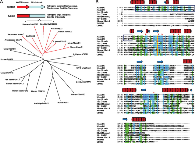
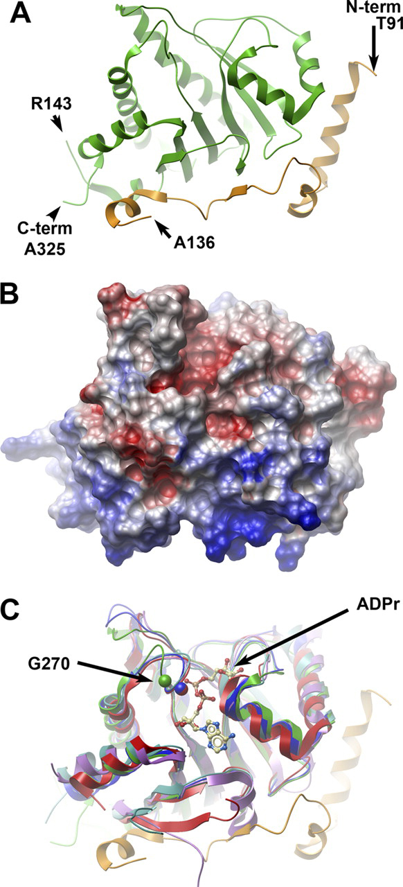
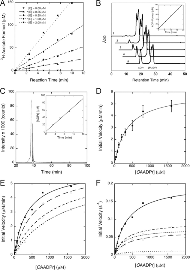
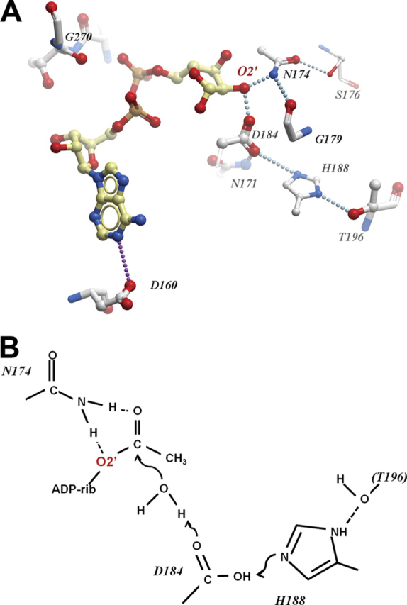
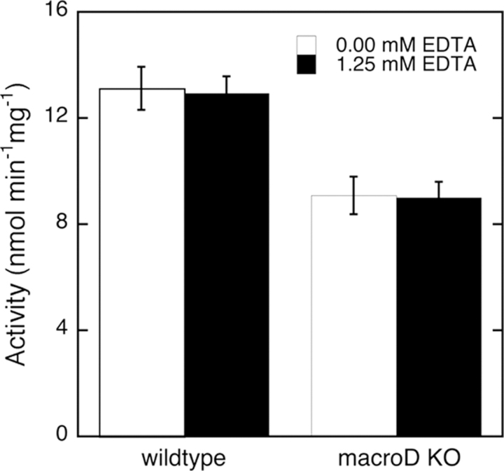
Sirtuins are a family of protein lysine deacetylases, which regulate gene silencing, metabolism, life span, and chromatin structure. Sirtuins utilize NAD(+) to deacetylate proteins, yielding O-acetyl-ADP-ribose (OAADPr) as a reaction product. The macrodomain is a ubiquitous protein module known to bind ADP-ribose derivatives, which diverged through evolution to support many different protein functions and pathways. The observation that some sirtuins and macrodomains are physically linked as fusion proteins or genetically coupled through the same operon, provided a clue that their functions might be connected. Indeed, here we demonstrate that the product of the sirtuin reaction OAADPr is a substrate for several related macrodomain proteins: human MacroD1, human MacroD2, Escherichia coli YmdB, and the sirtuin-linked MacroD-like protein from Staphylococcus aureus. In addition, we show that the cell extracts derived from MacroD-deficient Neurospora crassa strain exhibit a major reduction in the ability to hydrolyze OAADPr. Our data support a novel function of macrodomains as OAADPr deacetylases and potential in vivo regulators of cellular OAADPr produced by NAD(+)-dependent deacetylation.


Figures

FIGURE 1
FIGURE 1
MacroD-like proteins as a distinct subfamily of macrodomain proteins that includes sirtuin-linked factors.A, phylogenetic tree illustrating relationships between different macrodomain proteins. Branch of MacroD-like proteins is shown in red. Schematic representation of the genome arrangements of sirtuin-linked macrodomain proteins is shown in the box, and it relates to MacroD protein from C. albicans and SAV0325 protein from S. aureus shown in the tree. B, structure-based alignment of macrodomain proteins. The protein chains were aligned according to three-dimensional structures. A sequence-based alignment (ClustalW) was then manually adjusted to align spatially equivalent residues. The boxed regions are the N-terminal segments common to human MacroD1 and MacroD2 but absent from other macrodomain proteins; the red lettering above the sequences indicate residues which, when mutated, resulted in reduced enzymatic activity. The cylinders and arrows above the sequences represent, respectively, helices and β-strands in the structure of MacroD1. The proteins are: MacroD1 (PDB code 2X47, this study); MacroD2 (sequence only), E. coli YmdB (PDB code 1SPV); Yeast YMX7 (1TXZ)(37); Thermus thermophilus hypothetical protein (2DX6); SARS virus (2FAV)(4); Human histone H2A1.1 (3IIF)(36), human PARP15 macrodomain (3KH6), and human GDAP2 (sequence only).
FIGURE 2
FIGURE 2
The crystal structure of human MacroD1.A, ribbon representation of the secondary structure of MacroD1. The conserved macrodomain is depicted in green, and the divergent N-terminal (aa 91–136) is in orange. The amino acids at the two termini of the ordered structure are marked, as well as the breakpoints of an internally missing fragment. B, surface potential diagram of MacroD1 shown in the same orientation as Fig. 2A. The N-terminal region (bottom) is highly positive. C, overlay of MacroD1 (green/orange) on the structures of other macrodomains: Feline sarcoma virus (PDB code 3JZT; purple), E. coli YmdB (PDB code 1SPV; blue), human PARP15 (PDB code 3KH6; cyan), and histone macroH2A1.1 (PDB code 3IID; red). An ADPr molecule associated with the Feline sarcoma virus protein is shown as an atomic model. The Cα atom of a conserved glycine (Gly-270 of MacroD1) is marked in a green sphere; the homologous glycine of YmdB and MacroH2A1.1 are marked with blue and red spheres, respectively. The shift of position of the loop containing Gly-270 (green) in MacroD1 relative to all other macrodomain structures is most likely the result of a close contact with a neighboring molecule in the crystal involving residues Val-271 and Phe-272 (not shown).
FIGURE 3
FIGURE 3
Deacetylation of O-acetyl-ADPr catalyzed by macroD-like proteins.A, [3H]acetate release from hydrolysis of O-[3H]acetyl-ADPr catalyzed by human MacroD1. OAADPr hydrolysis was monitored by measuring the radioactivity release as count per minute (CPM) due to [3H]acetate formation from O-[3H]acetyl-ADPr in the presence of 0 to 2.0 μm human MacroD1 and 800 μmOAADPr at pH 7.3. The data show the time dependent increase in free [3H]acetate formation and the rate of [3H]acetate formation increases with Macro D1 (E) concentration. B, HPLC assay of OAADPr hydrolysis catalyzed by human MacroD1. HPLC chromatograms monitored at 260 nm show the formation of ADPr from OAADPr in human MacroD1 catalyzed reaction. The chromatograms labeled 1–5 are reactions stopped by addition of TFA at 0, 3, 6, 10, and 16 min, respectively. The formation of ADPr from OAADPr depends on the presence of enzyme. Inset shows the time-dependent formation of ADPr from OAADPr in the presence of 0.5 μm MacroD1. C, LC-MS/MS analysis of human MacroD1 reaction. The precursor ions of 600.0 (OAADPr) and 558.0 (ADPr) and the major product ion of 346.0 shared by OAADPr and ADPr were selected. The SRM chromatogram shows the ion intensity versus time for OAADPr (dashed line) and ADPr (solid line) formed from the MacroD1 reaction stopped at 15 min. The inset shows the formation of ADPr increases with reaction time. The concentration of ADPr formed from each time point reaction was determined from external standards. D, steady-state kinetic analysis of human MacroD1. The deacetylation reaction of OAADPr catalyzed by MacroD1 follows saturation kinetics. The steady-state kinetic parameters are determined by radioactive assays for acetate formation and HPLC assays for ADP-ribose formation. An apparent Km was measured to be 375 ± 55 μm. The Vmax was measured to be 0.20 ± 0.04 s−1. The reaction mixtures contain 0.5 μm MacroD1. E, inhibition of ADPr to human MacroD1 activity. The deacetylation reactions were carried out in 50 mm Tris-HCl of pH 7.3 containing 0.5 μm MacroD1. The reactions were initiated in the presence of 0 μm (closed squares), 100 μm (open squares), 200 μm (open diamonds), and 400 μm (open circles) ADPr, respectively. The Km′ values measured are 375.8, 590.9, 986.6, and 1350.3 μm in the presence of 0, 100, 200, and 400 μm initial ADPr, respectively. The Vmax values are 0.202, 0.204, 0.201, and 0.196 s−1. The Ki was determined to be 145 ± 27 μm. The inhibition pattern is consistent with reversible competitive inhibition. F, kinetic comparison of wild-type MacroD1 and its variants. Conserved asparagine and aspartate residues located in vicinity to the 2′- and 3′-hydroxyl groups of the bound ADPr in the MacroD1 structure are mutated to alanine. The kinetic comparison of the wild type MacroD1 (closed squares) and its variants show that these residues are important to catalysis. The N171A (open squares), N174A (open diamonds), D184A (open circles), and N174A/D184A (open triangles), mainly affect the Vmax. The Km values are only minimally affected.
FIGURE 4
FIGURE 4
Possible arrangement of the MacroD1 catalytic site.A, residues of MacroD1 surrounding the modeled ADPr binding site. Dotted lines indicate potential hydrogen bonds either direct or water-mediated. B, hypothetical roles of catalytic residues in the hydrolysis of OAADPr.
FIGURE 5
FIGURE 5
OAADPr deacetylation activity of cell extracts from wild type and MacroD knock-out (Δmacrod) neurospora.Neurospora cell extracts were assayed using radioactive method in the absence or presence of 1.25 mm EDTA. Protein concentrations in cell extracts were normalized before activity assays. MacroD1 knock-out in Neurospora reduced the OAADPr deacetylation activities in cell extracts for ∼30% independent of EDTA. The error bars represent standard deviations calculated from the specific activities of Neurospora extracts in five separate experiments.

References

    1. Till S., Ladurner A.G. Front. Biosci. 2009;14:3246–3258. - PubMed
    1. Ladurner A.G. Mol. Cell. 2003;12:1–3. - PubMed
    1. Karras G.I., Kustatscher G., Buhecha H.R., Allen M.D., Pugieux C., Sait F., Bycroft M., Ladurner A.G. EMBO J. 2005;24:1911–1920. - PMC - PubMed
    1. Egloff M.P., Malet H., Putics A., Heinonen M., Dutartre H., Frangeul A., Gruez A., Campanacci V., Cambillau C., Ziebuhr J., Ahola T., Canard B. J. Virol. 2006;80:8493–8502. - PMC - PubMed
    1. Neuvonen M., Ahola T. J. Mol. Biol. 2009;385:212–225. - PMC - PubMed

Publication types

MeSH terms

Associated data