

Articular cartilage in the knee: current MR imaging techniques and applications in clinical practice and research
- PMID: 21257932
- PMCID: PMC6939857
- DOI: 10.1148/rg.311105084
Articular cartilage in the knee: current MR imaging techniques and applications in clinical practice and research
Abstract
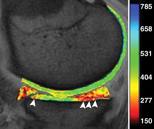
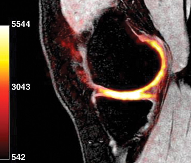
Magnetic resonance (MR) imaging is the most important imaging modality for the evaluation of traumatic or degenerative cartilaginous lesions in the knee. It is a powerful noninvasive tool for detecting such lesions and monitoring the effects of pharmacologic and surgical therapy. The specific MR imaging techniques used for these purposes can be divided into two broad categories according to their usefulness for morphologic or compositional evaluation. To assess the structure of knee cartilage, standard spin-echo (SE) and gradient-recalled echo (GRE) sequences, fast SE sequences, and three-dimensional SE and GRE sequences are available. These techniques allow the detection of morphologic defects in the articular cartilage of the knee and are commonly used in research for semiquantitative and quantitative assessments of cartilage. To evaluate the collagen network and proteoglycan content in the knee cartilage matrix, compositional assessment techniques such as T2 mapping, delayed gadolinium-enhanced MR imaging of cartilage (or dGEMRIC), T1ρ imaging, sodium imaging, and diffusion-weighted imaging are available. These techniques may be used in various combinations and at various magnetic field strengths in clinical and research settings to improve the characterization of changes in cartilage.
RSNA, 2011
Figures


Comment in
-
Articular cartilage in the knee: current MR imaging techniques and applications in clinical practice and research. Invited commentary.Radiographics. 2011 Jan-Feb;31(1):61-2. doi: 10.1148/Radiographics.31.1.31161. Radiographics. 2011. PMID: 21322827 No abstract available.
-
Potential use of pharmacokinetic modeling of first-pass dynamic contrast-enhanced MR images of cartilage.Radiographics. 2011 May-Jun;31(3):894-5. doi: 10.1148/Radiographics.31.3.313894. Radiographics. 2011. PMID: 21571666 No abstract available.
References
-
- Felson DT. Osteoarthritis of the knee. N Engl J Med 2006;354(8):841–848. - PubMed
-
- Gold GE, McCauley TR, Gray ML, Disler DG. What's new in cartilage? RadioGraphics 2003;23(5):1227–1242. - PubMed
-
- Burstein D, Gray M, Mosher T, Dardzinski B. Measures of molecular composition and structure in osteoarthritis. Radiol Clin North Am 2009;47(4):675–686. - PubMed
-
- Trattnig S, Domayer S, Welsch GW, Mosher T, Eckstein F. MR imaging of cartilage and its repair in the knee: a review. Eur Radiol 2009;19(7):1582–1594. - PubMed
Publication types
MeSH terms
Grants and funding
LinkOut - more resources
Full Text Sources
Other Literature Sources
Medical
