Comparative Study
. 2011 Feb 15;186(4):2106-16.
doi: 10.4049/jimmunol.1002000. Epub 2011 Jan 21.

CD4 and CD8 T cell immune activation during chronic HIV infection: roles of homeostasis, HIV, type I IFN, and IL-7

Affiliations
Comparative Study

CD4 and CD8 T cell immune activation during chronic HIV infection: roles of homeostasis, HIV, type I IFN, and IL-7

Marta Catalfamo et al. J Immunol. .

Abstract

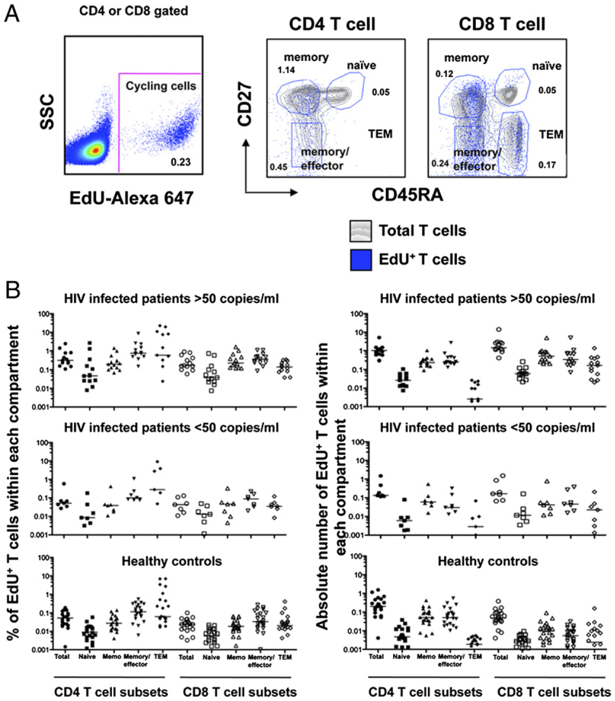
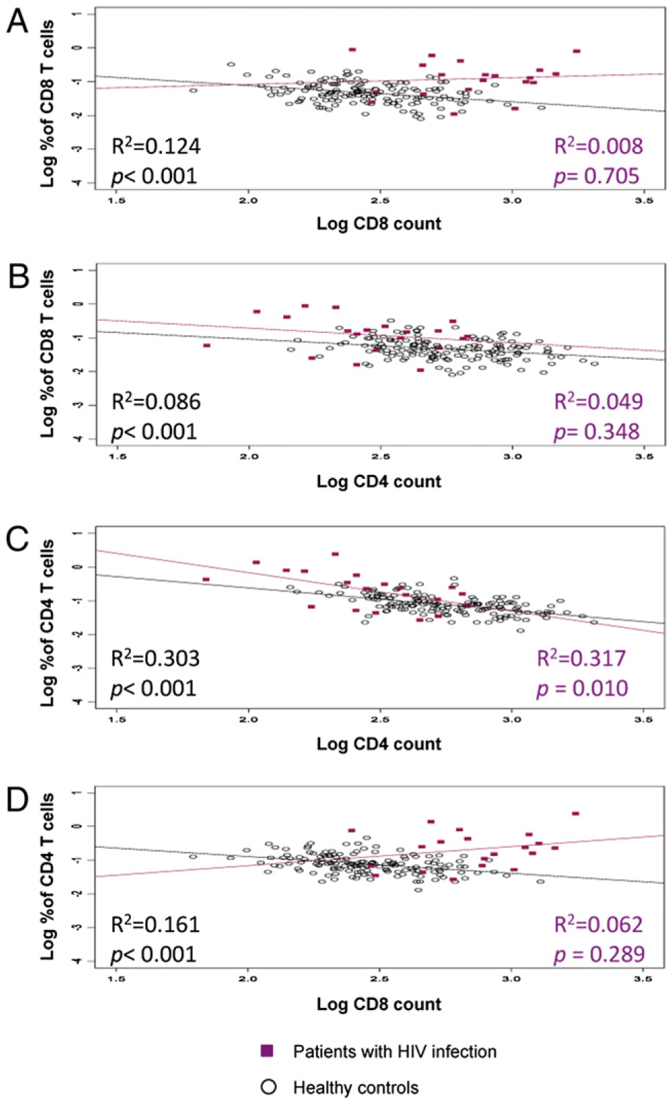
Immune activation plays an important role in the pathogenesis of HIV disease. Although the causes are not fully understood, the forces that lead to immune dysfunction differ for CD4 and CD8 T cells. In this study, we report that the molecular pathways that drive immune activation during chronic HIV infection are influenced by differences in the homeostatic regulation of the CD4 and CD8 T cell pools. Proliferation of CD4 T cells is controlled more tightly by CD4 T cell numbers than is CD8 T cell proliferation. This difference reflects the importance of maintaining a polyclonal CD4 T cell pool in host surveillance. Both pools of T cells were found to be driven by viral load and its associated state of inflammation. In the setting of HIV-induced lymphopenia, naive CD4 T cells were recruited mainly into the proliferating pool in response to CD4 T cell depletion, whereas naive CD8 T cell proliferation was driven mainly by levels of HIV RNA. RNA analysis revealed increased expression of genes associated with type I IFN and common γ chain cytokine signaling in CD4 T cell subsets and only type I IFN-associated genes in CD8 T cell subsets. In vitro studies demonstrated enhanced STAT1 phosphorylation in response to IFN-α and increased expression of the IFNAR1 transcripts in naive and memory CD4 T cells compared with that observed in CD8 T cells. CD4 T cell subsets also showed enhanced STAT1 phosphorylation in response to exogenous IL-7.


Conflict of interest statement

Disclosures

The authors have no financial conflicts of interest.

Figures

FIGURE 1.
FIGURE 1.
Ex vivo proliferation of CD4 and CD8 T cell subsets determined by EdU incorporation. A, PBMCs from patients were labeled with EdU to measure spontaneous ex vivo proliferation of T cell subsets: naive (CD45RA+CD27+), memory (CD45RACD27+), effector/memory (CD45RACD27), and TEM (CD45RA+CD27). The gray contour plots represent the total population of CD4 or CD8 T cells, and the overlay blue dot plot represents the proliferating population. The percentages indicate the percentages of the cells within each gate that are cycling. B, Percentages and the absolute numbers of proliferating CD4 and CD8 T cell subsets from patients with HIV infection and HIV RNA levels of <50 or >50 copies/ml) (Table I). Bars in the graph represent median values. Actual numbers for the medians are presented in Table I.
FIGURE 2.
FIGURE 2.
Relationships between CD4 or CD8 proliferating T cells and CD4 or CD8 T cell counts. Ex vivo proliferation of T cells depicted from healthy controls (n = 152, black dots) and HIV-infected individuals (n = 20, red squares). The association between percentage of CD8 T cells proliferating and CD8 T cell counts (A) or CD4 T cell counts (B) and the association of percentage of CD4 T cells proliferating and CD4 T cell counts (C) or CD8 T cell counts (D).
FIGURE 3.
FIGURE 3.
Changes in CD4 and CD8 T cell counts and proliferation during ART. Ex vivo BrdU labeling at several time points was performed in patients (n = 36) undergoing ART. A and B, Two patients, one with a decrease in CD8 T cell count (A) and one with an increase in CD8 T cell count (B). C, Comparison of patients where CD8 T cell count increased significantly (n = 6) and decreased significantly (n = 4) after ART. Relationships with baseline CD4 and CD8 T cell count and viral load are shown.
FIGURE 4.
FIGURE 4.
STAT1 and STAT5 phosphorylation in response to in vitro stimulation with IFN-α, IL-7, and a combination of both cytokines. PBMCs from healthy volunteers (n = 10), HIV-infected individuals with HIV RNA levels of <50 copies/ml (n = 11), and HIV-infected individuals with HIV RNA levels of >30,000 copies/ml (n = 10) were stimulated in vitro with IL-7 (1 ng/ml), IFN-α (100 U/ml), or a combination of both cytokines. Phosphorylation of STAT1 and STAT5 in naive (CD45RA+CD27+) (A) and memory (CD45RA+CD27+) (B) T cell subsets was analyzed by multicolor flow cytometry in CD4 and CD8 T cells. The Mann-Whitney U test was performed for the comparison between groups.

References

    1. Sachsenberg N, Perelson AS, Yerly S, Schockmel GA, Leduc D, Hirschel B, and Perrin L. 1998. Turnover of CD4+ and CD8+ T lymphocytes in HIV-1 infection as measured by Ki-67 antigen. J. Exp. Med 187: 1295–1303. - PMC - PubMed
    1. Ho DD, Neumann AU, Perelson AS, Chen W, Leonard JM, and Markowitz M. 1995. Rapid turnover of plasma virions and CD4 lymphocytes in HIV-1 infection. Nature 373: 123–126. - PubMed
    1. Kovacs JA, Lempicki RA, Sidorov IA, Adelsberger JW, Herpin B, Metcalf JA, Sereti I, Polis MA, Davey RT, Tavel J, et al. 2001. Identification of dynamically distinct subpopulations of T lymphocytes that are differentially affected by HIV. J. Exp. Med 194: 1731–1741. - PMC - PubMed
    1. Lempicki RA, Kovacs JA, Baseler MW, Adelsberger JW, Dewar RL, Natarajan V, Bosche MC, Metcalf JA, Stevens RA, Lambert LA, et al. 2000. Impact of HIV-1 infection and highly active antiretroviral therapy on the kinetics of CD4+ and CD8+ T cell turnover in HIV-infected patients. Proc. Natl. Acad. Sci. USA 97: 13778–13783. - PMC - PubMed
    1. Hazenberg MD, Hamann D, Schuitemaker H, and Miedema F. 2000. T cell depletion in HIV-1 infection: how CD4+ T cells go out of stock. Nat. Immunol 1: 285–289. - PubMed

Publication types

MeSH terms

Associated data