. 2011 Mar 1;186(5):3113-9.
doi: 10.4049/jimmunol.1003189. Epub 2011 Jan 21.

A novel human IgA monoclonal antibody protects against tuberculosis

Affiliations

A novel human IgA monoclonal antibody protects against tuberculosis

Sucharitha Balu et al. J Immunol. .

Abstract

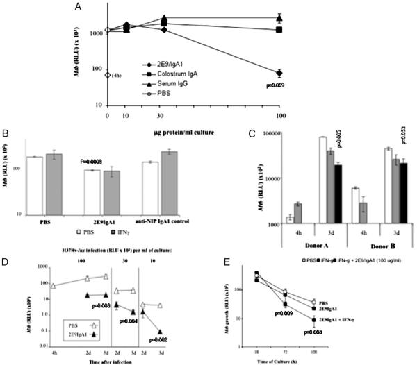
Abs have been shown to be protective in passive immunotherapy of tuberculous infection using mouse experimental models. In this study, we report on the properties of a novel human IgA1, constructed using a single-chain variable fragment clone (2E9), selected from an Ab phage library. The purified Ab monomer revealed high binding affinities for the mycobacterial α-crystallin Ag and for the human FcαRI (CD89) IgA receptor. Intranasal inoculations with 2E9IgA1 and recombinant mouse IFN-γ significantly inhibited pulmonary H37Rv infection in mice transgenic for human CD89 but not in CD89-negative littermate controls, suggesting that binding to CD89 was necessary for the IgA-imparted passive protection. 2E9IgA1 added to human whole-blood or monocyte cultures inhibited luciferase-tagged H37Rv infection although not for all tested blood donors. Inhibition by 2E9IgA1 was synergistic with human rIFN-γ in cultures of purified human monocytes but not in whole-blood cultures. The demonstration of the mandatory role of FcαRI (CD89) for human IgA-mediated protection is important for understanding of the mechanisms involved and also for translation of this approach toward development of passive immunotherapy of tuberculosis.


Figures

FIGURE 1
FIGURE 1
Characterization of purified 2E9IgA1. SDS-PAGE (Coomassie) and Western blot analysis probed with anti-L chain, anti-IgA, Con A, jacalin (Jac), Acr followed by anti-Acr, and Fc-(CD89)2. Serum IgA served as a control for Fc-(CD89)2 binding. M, m.w. markers; NR, non-reduced samples; R, reduced samples.
FIGURE 2
FIGURE 2
Modulation of M. tuberculosis infection in FcαRI/CD89 transgenic mice. Mice in all three groups were preinoculated i.n. with 1 μg mouse IFN-γ 3 d before i.n. infection with 0.5 million H37Rv. Five micrograms purified 2E9IgA1 (titer 16,500) mixed with either IFN-γ (closed symbols) or PBS (open symbols) was given i.n. 2 h before infection and again either 1 d or 21 d postinfection. Organs were harvested 4 wk postinfection. A, Group geometric means (horizontal bars) of CFU counts in the lungs (circles) and spleens (triangles) of individual mice. **p < 0.001, *p < 0.05 (significant difference between 2E9IgA1-inoculated and PBS-inoculated groups; t test). B, H&E-stained lung sections. Original magnification ×20. C, Means ± SE and t test values of granulomatous infiltration of the lungs from CD89tg mice. **p < 0.001.
FIGURE 3
FIGURE 3
Influence of combined and single i.n. inoculations of 2E9IgA1 and IFN-γ on M. tuberculosis infection. One microgram mouse IFN-γ was given 3 d before infection with 0.5 million H37Rv. Five micrograms purified 2E9IgA1 mixed with either IFN-γ or PBS was given 2 h before infection and again 1 d and 21 d postinfection. Organs were harvested 4 wk postinfection. CFU count data in the lungs (circles) and spleens (triangles) of individual mice and group geometric means (horizontal bars) are shown. *p < 0.05 (significant difference compared with the PBS-inoculated control group; t test).
FIGURE 4
FIGURE 4
Modulation of H37Rv-lux infection in vitro. Whole human blood cultures were incubated with 10 ng/ml human IFN-γ and 2E9IgA1 for 24 h prior to infection with 106 RLU H37Rv-lux for 2 h, followed by amikacin (200 μg/ml) treatment for 4 h. Chemiluminescence was determined after incubation at 37°C for 3 d. A, Influence of 2E9IgA, colostrum IgA, and serum IgG dose. B and C, Influence of human IFN-γ in different donors. D, Influence of different dosage of H37Rv-lux infection using 2E9IgA1 (closed symbols) or PBS (open symbols). E, Modulation of infection of purified human monocytes with 20 μg/ml 2E9IgA1 with or without IFN-γ before infection with H37Rv-lux. The p values represent the significance of the difference in comparison with the PBS controls (t test).

References

    1. Nunn P, Williams B, Floyd K, Dye C, Elzinga G, Raviglione M. Tuberculosis control in the era of HIV. Nat. Rev. Immunol. 2005;5:819–826. - PubMed
    1. Glatman-Freedman A, Casadevall A. Serum therapy for tuberculosis revisited: reappraisal of the role of antibody-mediated immunity against Mycobacterium tuberculosis. Clin. Microbiol. Rev. 1998;11:514–532. - PMC - PubMed
    1. Teitelbaum R, Glatman-Freedman A, Chen B, Robbins JB, Unanue E, Casadevall A, Bloom BR. A mAb recognizing a surface antigen of Mycobacterium tuberculosis enhances host survival. Proc. Natl. Acad. Sci. USA. 1998;95:15688–15693. - PMC - PubMed
    1. Hamasur B, Haile M, Pawlowski A, Schroder U, Kallenius G, Svenson SB. A mycobacterial lipoarabinomannan specific monoclonal antibody and its F(ab’) fragment prolong survival of mice infected with Mycobacterium tuberculosis. Clin. Exp. Immunol. 2004;138:30–38. - PMC - PubMed
    1. Pethe K, Alonso S, Biet F, Delogu G, Brennan MJ, Locht C, Menozzi FD. The heparin-binding haemagglutinin of M. tuberculosis is required for extrapulmonary dissemination. Nature. 2001;412:190–194. - PubMed

Publication types

MeSH terms