. 2011 Mar;22(3):526-35.
doi: 10.1681/ASN.2010050522. Epub 2011 Jan 21.

TRPC6 enhances angiotensin II-induced albuminuria

Affiliations

TRPC6 enhances angiotensin II-induced albuminuria

Jason Eckel et al. J Am Soc Nephrol. 2011 Mar.

Abstract

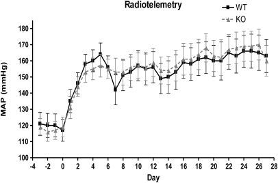
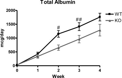
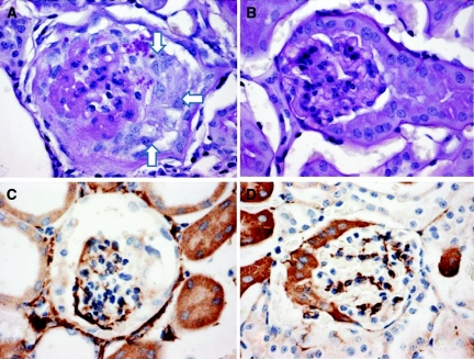
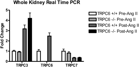
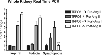
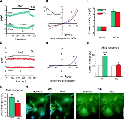
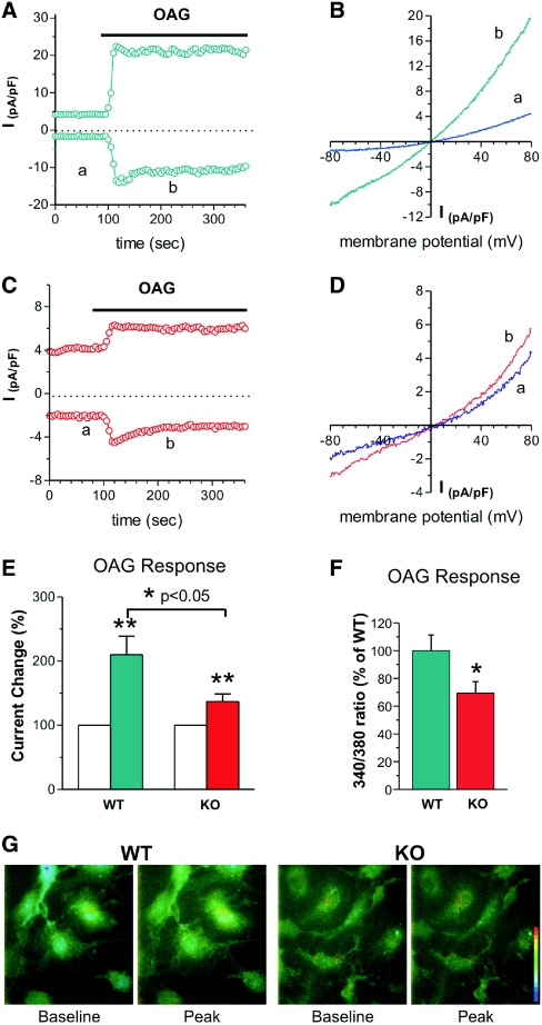
Mutations in the canonical transient receptor potential cation channel 6 (TRPC6) are responsible for familial forms of adult onset focal segmental glomerulosclerosis (FSGS). The mechanisms by which TRPC6 mutations cause kidney disease are not well understood. We used TRPC6-deficient mice to examine the function of TRPC6 in the kidney. We found that adult TRPC6-deficient mice had BP and albumin excretion rates similar to wild-type animals. Glomerular histomorphology revealed no abnormalities on both light and electron microscopy. To determine whether the absence of TRPC6 would alter susceptibility to hypertension and renal injury, we infused mice with angiotensin II continuously for 28 days. Although both groups developed similar levels of hypertension, TRPC6-deficient mice had significantly less albuminuria, especially during the early phase of the infusion; this suggested that TRPC6 adversely influences the glomerular filter. We used whole-cell patch-clamp recording to measure cell-membrane currents in primary cultures of podocytes from both wild-type and TRPC6-deficient mice. In podocytes from wild-type mice, angiotensin II and a direct activator of TRPC6 both augmented cell-membrane currents; TRPC6 deficiency abrogated these increases in current magnitude. Our findings suggest that TRPC6 promotes albuminuria, perhaps by promoting angiotensin II-dependent increases in Ca(2+), suggesting that TRPC6 blockade may be therapeutically beneficial in proteinuric kidney disease.


Figures

Figure 1.
Figure 1.
Similar mean arterial pressure (MAP) in WT and TRPC6 knockout/null mice. MAP was measured by radiotelemetry. The solid line indicates WT (TRPC6+/+), n = 5; the dashed line shows KO (TRPC6−/−), n = 6. P = NS at all time points.
Figure 2.
Figure 2.
Trend toward decreased 24-hour albuminuria in knockout compared with wild-type mice. There is a significant difference between wild type and knockout at weeks 2 and 3 of angiotensin II infusion. There was less albuminuria in the KO mice at week 4; however, it was not statistically significant. For WT, n = 14; for KO, n = 18. #P = 0.004; ##P = 0.04.
Figure 3.
Figure 3.
Examples of glomerular damage after treatment with angiotensin II. (A and C) Glomerular sclerosis and collapse (white arrows in A) and increased desmin staining in podocytes (C). (B) Normal glomerular staining. (D) Normal desmin staining.
Figure 4.
Figure 4.
Upregulation of TRPCs 3 and 6 and downregulation of TRPC7 after angiotensin II treatment. Real-time PCR was used to evaluate mouse kidney tissue. (+/+ is TRPC6 wild type and −/− is TRPC6 knockout).
Figure 5.
Figure 5.
Upregulation of nephrin and podocin and downregulation of synaptopodin in knockout mice (TRPC6 −/−) after angiotensin II treatment. Real-time PCR was used to evaluate mouse kidney tissue. **P < 0.01 in knockout (−/−) versus wild type (TRPC6 +/+).
Figure 6.
Figure 6.
Reduced angiotensin II-generated membrane currents (INSC) and Ca2+ signals in TRPC6 KO podocytes. (A) An example of membrane current recorded from a WT podocyte. The current was induced by a 200-ms voltage ramp protocol (1 mV/ms, from 100 mV to −100 mV, holding potential 0 mV, see inset) every 3 seconds. INSC was normalized by membrane capacitance. The current measured at −80 mV. Note that the current was increased after Ang II (ANG, 10 μM) perfusion and inhibited by gadolinium (Gd, 10 μM). (B) Examples of I/V relation of membrane currents in the WT podocyte. The letters a through c correspond to the traces in A. (C) An example of membrane current recorded from a TRPC6 KO podocyte. The recording protocol was same as above. Note that the current was not increased by Ang II. (D) Examples of I/V relation of membrane currents in the KO podocyte. The letters d through f correspond to the traces in C. (E) Group mean values of INSC at −80 mV and +80 mV in WT (green bars, n = 29) and TRPC6 KO podocytes (red bars, n = 22). There is no significant difference between WT and KO podocytes. (F) Group mean changes (%) of INSC at −80 mV caused by perfusion of Ang II (ANG 10 μM) in WT and TRPC6 KO podocytes. Ang II nearly doubled INSC in WT podocytes (green bar, n = 16, **P < 0.01), whereas Ang II slightly increased INSC in TRPC6 KO podocytes (red bar, n = 23, *P < 0.05). (G) Group mean Ca2+ increases (% of WT) produced by Ang II (100 μM) in WT (green bar, n = 32) and TRPC6 KO (red bar, n = 47) podocytes. The magnitude of the TRPC6 KO Ca2+ response is 69.9 ± 0.085% of the WT response (*P < 0.05). (H) Examples of Ca2+ signals produced by Ang II in WT and KO podocytes. The pseudocolor scale (bar) is from 340/380 ratio 0.1 to 1.0. Note that ANG produced a larger Ca2+ increase in WT than in KO podocytes.
Figure 7.
Figure 7.
Membrane currents (INSC) and Ca2+ signals in response to OAG stimulation are reduced in TRPC6 KO podocytes. (A) An example of membrane current recorded from a WT podocyte. The current was recorded as described above. The current measured at −80 mV and +80 mV. Note that the current was increased after OAG (100 μM) perfusion. (B) Examples of I/V relation of membrane currents in the WT podocyte. The letters a and b correspond to the traces in A. (C) An example of membrane current recorded from a TRPC6 KO podocyte. The recording protocol was the same as above. Note that the current was also increased by OAG but less than that in WT cells. (D) Examples of I/V relation of membrane currents in the KO podocyte. The letters a and b correspond to the traces in C. (E) Group mean changes (%) of INSC at −80 mV caused by OAG (100 μM) in WT and TRPC6 KO podocytes. OAG doubled INSC in WT podocytes (green bar, n = 16; **P < 0.01), whereas the increase was attenuated by 37% in TRPC6 KO podocytes (red bar, n = 23; *P < 0.05). (F) Group mean Ca2+ increases (% of WT) produced by OAG (100 μM) in WT (green bar, n = 58) and TRPC6 KO (red bar, n = 59) podocytes. The magnitude of the TRPC6 KO response is 69.4 ± 0.082% of the WT response (*P < 0.05). (G) Examples of Ca2+ signals produced OAG in WT and KO podocytes. The pseudocolor scale (bar) is from 340/380 ratio 0.1 to 1.0. Note that OAG produced a larger Ca2+ increase in WT than in KO podocytes.

References

    1. Dietrich A, Kalwa H, Rost BR, Gudermann T: The diacylgylcerol-sensitive TRPC3/6/7 subfamily of cation channels: Functional characterization and physiological relevance. Pflugers Arch 451: 72–80, 2005 - PubMed
    1. Winn MP, Conlon PJ, Lynn KL, Farrington MK, Creazzo T, Hawkins AF, Daskalakis N, Kwan SY, Ebersviller S, Burchette JL, Pericak-Vance MA, Howell DN, Vance JM, Rosenberg PB: A mutation in the TRPC6 cation channel causes familial focal segmental glomerulosclerosis. Science 308: 1801–1804, 2005 - PubMed
    1. Kestila M, Lenkkeri U, Mannikko M, Lamerdin J, McCready P, Putaala H, Ruotsalainen V, Morita T, Nissinen M, Herva R, Kashtan CE, Peltonen L, Holmberg C, Olsen A, Tryggvason K: Positionally cloned gene for a novel glomerular protein–nephrin–is mutated in congenital nephrotic syndrome. Mol Cell 1: 575–582, 1998 - PubMed
    1. Boute N, Gribouval O, Roselli S, Benessy F, Lee H, Fuchshuber A, Dahan K, Gubler MC, Niaudet P, Antignac C: NPHS2, encoding the glomerular protein podocin, is mutated in autosomal recessive steroid-resistant nephrotic syndrome. Nat Genet 24: 349–354, 2000 - PubMed
    1. Shankland SJ: The podocyte's response to injury: Role in proteinuria and glomerulosclerosis. Kidney Int 69: 2131–2147, 2006 - PubMed

Publication types

MeSH terms

LinkOut - more resources