. 2011 Apr 8;286(14):11937-50.
doi: 10.1074/jbc.M110.203661. Epub 2011 Jan 23.

Reversible high affinity inhibition of phosphofructokinase-1 by acyl-CoA: a mechanism integrating glycolytic flux with lipid metabolism

Affiliations

Reversible high affinity inhibition of phosphofructokinase-1 by acyl-CoA: a mechanism integrating glycolytic flux with lipid metabolism

Christopher M Jenkins et al. J Biol Chem. .

Abstract

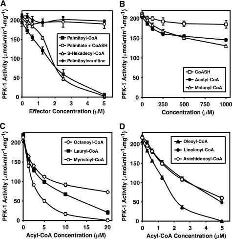
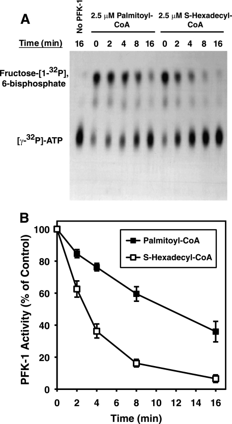
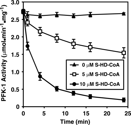
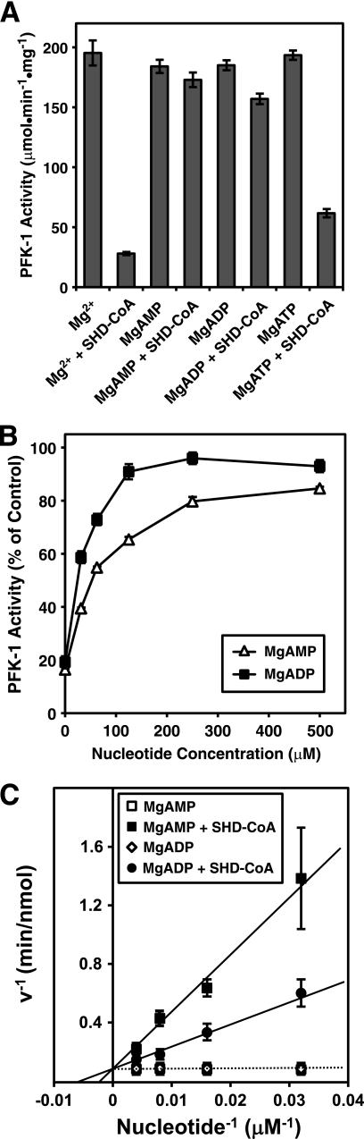
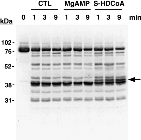
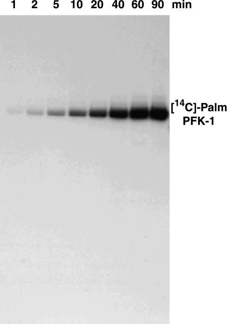
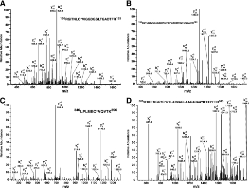
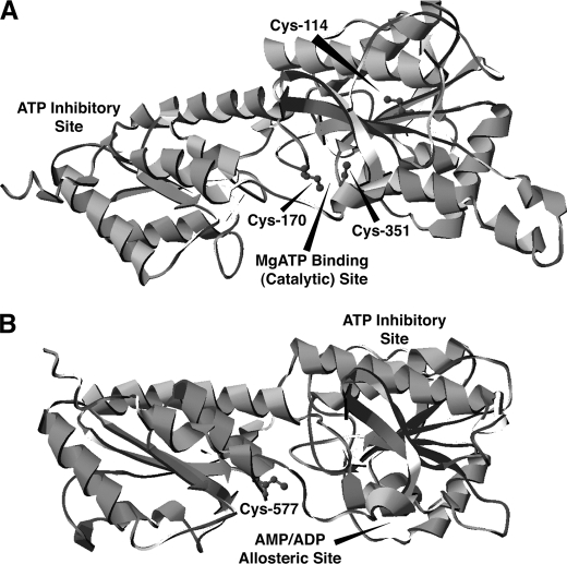
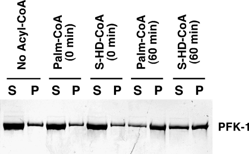
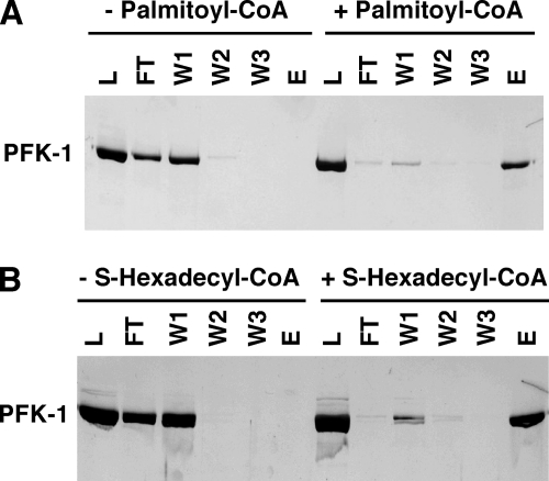
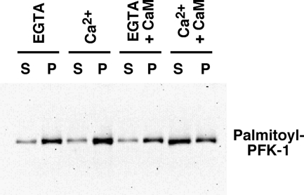
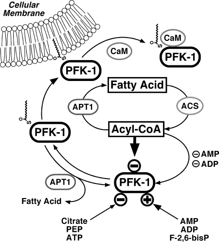
The enzyme phosphofructokinase-1 (PFK-1) catalyzes the first committed step of glycolysis and is regulated by a complex array of allosteric effectors that integrate glycolytic flux with cellular bioenergetics. Here, we demonstrate the direct, potent, and reversible inhibition of purified rabbit muscle PFK-1 by low micromolar concentrations of long chain fatty acyl-CoAs (apparent Ki∼1 μM). In sharp contrast, short chain acyl-CoAs, palmitoylcarnitine, and palmitic acid in the presence of CoASH were without effect. Remarkably, MgAMP and MgADP but not MgATP protected PFK-1 against inhibition by palmitoyl-CoA indicating that acyl-CoAs regulate PFK-1 activity in concert with cellular high energy phosphate status. Furthermore, incubation of PFK-1 with [1-(14)C]palmitoyl-CoA resulted in robust acylation of the enzyme that was reversible by incubation with acyl-protein thioesterase-1 (APT1). Importantly, APT1 reversed palmitoyl-CoA-mediated inhibition of PFK-1 activity. Mass spectrometric analyses of palmitoylated PFK-1 revealed four sites of acylation, including Cys-114, Cys-170, Cys-351, and Cys-577. PFK-1 in both skeletal muscle extracts and in purified form was inhibited by S-hexadecyl-CoA, a nonhydrolyzable palmitoyl-CoA analog, demonstrating that covalent acylation of PFK-1 was not required for inhibition. Tryptic footprinting suggested that S-hexadecyl-CoA induced a conformational change in PFK-1. Both palmitoyl-CoA and S-hexadecyl-CoA increased the association of PFK-1 with Ca2+/calmodulin, which attenuated the binding of palmitoylated PFK-1 to membrane vesicles. Collectively, these results demonstrate that fatty acyl-CoA modulates phosphofructokinase activity through both covalent and noncovalent interactions to regulate glycolytic flux and enzyme membrane localization via the branch point metabolic node that mediates lipid flux through anabolic and catabolic pathways.


Figures

FIGURE 1.
FIGURE 1.
Inhibition of PFK-1 by low micromolar concentrations of long chain fatty acyl-CoAs and the dependence of inhibition on acyl chain length. A, palmitoyl-CoA and the nonhydrolyzable analog S-hexadecyl-CoA, but not palmitic acid or CoASH alone or in combination, inhibit PFK-1 activity. Increasing concentrations of each reagent as indicated were incubated with purified rabbit skeletal muscle PFK-1 (0.1 μm) for 60 min at 30 °C in 100 mm Tris-HCl, pH 7.5, containing 1 mm DTT. Fructose-6-phosphate 1-kinase activity was measured at 22 °C using a coupled spectrophotometric assay as described under “Experimental Procedures.” B, effect of CoASH and short chain acyl-CoAs on PFK-1 activity. C, effect of medium chain length fatty acyl-CoAs on PFK-1 activity. D, effect of unsaturated fatty acyl-CoAs on phosphofructokinase activity. Increasing concentrations of fatty acyl-CoAs of varying chain length were incubated with purified PFK-1 and assayed as described above. Results are the mean ± S.E. from at least three separate experiments.
FIGURE 2.
FIGURE 2.
Time course of palmitoyl-CoA- and S-hexadecyl-CoA-mediated inhibition of PFK-1 activity utilizing a direct radiometric assay employing [γ-32P]ATP. Purified rabbit muscle PFK-1 (0.1 μm) was incubated with palmitoyl-CoA (2.5 μm) in 25 mm Tris-HCl, pH 7.5, containing 50 mm NaCl and 1 mm DTT for the indicated times. PFK activity was then assayed in the presence of 1 mm fructose 6-phosphate and 100 μm [γ-32P]ATP (20 μCi/μmol) for 1 min at 25 °C. After termination of the reaction by addition of acetone, the products were resolved by TLC, detected by autoradiography, and quantified by densitometry as described under “Experimental Procedures.” A, representative TLC autoradiograph showing inhibition of fructose [1-32P]6-bisphosphate formation in the absence and presence of palmitoyl-CoA or S-hexadecyl-CoA. B, quantitation of palmitoyl-CoA and S-hexadecyl-CoA mediated inhibition of PFK-1 from three separate experiments (mean ± S.E.) performed as described above.
FIGURE 3.
FIGURE 3.
Time course of inhibition of PFK-1 activity by S-hexadecyl-CoA in rabbit muscle cytosol. Isolated rabbit muscle cytosol (0.5 mg/ml) in 100 mm Tris-HCl, pH 7.5, containing 1 mm DTT was incubated with either 0, 5, or 10 μm S-hexadecyl-CoA for the indicated times at 30 °C. PFK-1 catalytic activity was then determined using a coupled spectrophotometric assay measuring the rate of NADH oxidation employing aldolase, triose-phosphate isomerase, and glycerol-3-phosphate dehydrogenase as described under “Experimental Procedures.” Results presented are the mean ± S.E. from at least three separate experiments.
FIGURE 4.
FIGURE 4.
Protection of PFK-1 from S-hexadecyl-CoA-mediated inhibition by MgAMP and MgADP but not MgATP. A, purified rabbit muscle PFK-1 (0.1 μm) was preincubated for 5 min at 30 °C in the presence of either AMP (1 mm), ADP (1 mm), or ATP (1 mm) in 100 mm Tris-HCl, pH 7.5, buffer containing 1 mm DTT and 1 mm MgCl2. S-Hexadecyl-CoA (SHD-CoA) was then added to a 10 μm concentration, and the enzyme was incubated for 5 min at 30 °C before measuring phosphofructokinase activity as described under “Experimental Procedures.” B, purified rabbit muscle PFK-1 (0.1 μm) was preincubated with the indicated concentration of MgAMP or MgADP as described above before addition of 10 μm S-hexadecyl-CoA, incubation at 30 °C for 5 min, and measurement of PFK activity. C, double-reciprocal plot indicating competition between MgAMP or MgADP binding and S-hexadecyl-CoA (5 μm)-mediated inhibition of PFK-1 (0.1 μm) following incubation at 30 °C for 5 min as determined by kinetic rate analysis. Results are the mean ± S.E. from at least three separate experiments.
FIGURE 5.
FIGURE 5.
S-Hexadecyl-CoA alters the susceptibility of PFK-1 to proteolysis as determined by tryptic footprinting. Following preincubation of purified rabbit muscle PFK-1 (1 μm, 85 μg/ml) in the presence or absence of 0.25 mm MgAMP or 2.5 μm S-hexadecyl-CoA, sequencing grade modified trypsin was added at a 1:40 w/w ratio and incubated for the indicated times. Trypsinolysis reactions were terminated by addition of 2× SDS-PAGE loading buffer and vortexing. PFK-1 tryptic fragments were resolved by SDS-PAGE, transferred to PVDF membranes, and probed with an antibody directed against the C terminus of PFK-1. Immunoreactive bands were detected by enhanced chemiluminescence using a protein A-horseradish peroxidase conjugate and visualized using a Kodak Image Station as described under “Experimental Procedures.” A novel 42-kDa tryptic peptide generated only in the presence of S-hexadecyl-CoA is indicated by the arrow. Results are representative of three separate experiments. CTL, control.
FIGURE 6.
FIGURE 6.
Time course of covalent acylation of PFK-1 with [14C]palmitoyl-CoA. Purified rabbit muscle PFK-1 (1 μm) was incubated with 10 μm [1-14C]palmitoyl-CoA at 35 °C in 25 mm Tris-HCl, pH 7.5, containing 50 mm KCl and 1 mm DTT for the indicated times. Samples were resolved by SDS-PAGE and visualized by autoradiography as described under “Experimental Procedures.” Results presented are representative of at least three separate experiments.
FIGURE 7.
FIGURE 7.
Identification of the sites of palmitoyl-CoA-mediated palmitoylation of phosphofructokinase by mass spectrometry. Purified rabbit skeletal muscle PFK-1 (10 μm) was incubated in the presence or absence of a stoichiometric amount of palmitoyl (Palm)-CoA in 25 mm Tris-HCl, pH 7.5, containing 50 mm KCl and 1 mm DTT for 60 min at 35 °C. Protein samples were precipitated, trypsinized, and processed as described under “Experimental Procedures.” Ion peaks corresponding to predicted palmitoylated tryptic peptides in the full mass scan were selected for product ion analysis. A, product ion mass spectrum of the ion at 2461.34 corresponding to the tryptic peptide (RGITNLC*VIGGDGSLTGADTFR) palmitoylated at Cys-114. B, product ion mass spectrum of the ion at 3609.80 corresponding to the tryptic peptide (SSYLNIVGLVGSIDNDFC*GTDMTIGTDSALR) palmitoylated at Cys-170. C, product ion mass spectrum of the ion at 1498.89 corresponding to the tryptic peptide (LPLMEC*VQVTK) palmitoylated at Cys-351. D, product ion mass spectrum of the ion at 4171.10 corresponding to the tryptic peptide (VFIIETMGGYC*GYLATMAGLAAGADAAYIFEEPFTIR) palmitoylated at Cys-577. Asterisks indicate the sites of palmitoylation. Mass to charge (m/z) ratios of the identified b and y fragment ions are as indicated.
FIGURE 8.
FIGURE 8.
Localization of the palmitoylated cysteine residues identified by mass spectrometry in a molecular model of PFK-1. The primary sequence of rabbit muscle PFK-1 was used in combination with the known crystal structures of three bacterial phosphofructokinases for energy minimization modeling using I-TASSER as described under “Experimental Procedures.” A, molecular model of the N-terminal domain of rabbit muscle PFK-1 (residues 1–389) demonstrates the close spatial proximity of residues Cys-114, Cys-170, and Cys-351 identified as palmitoyl thioesters by mass spectrometry to the catalytic MgATP-binding site. B, model of the C-terminal domain of rabbit muscle PFK-1 (residues 390–763) identifies the homologous position of the acylated residue at Cys-577 in comparison with the identified palmitoylated residues in the N-terminal domain and its close localization to the AMP/ADP allosteric site.
FIGURE 9.
FIGURE 9.
Reversal of palmitoyl (Palm)-CoA inhibition of PFK-1 and deacylation of the enzyme by APT1. A, time course of [14C]palmitoyl-PFK deacylation by APT1. [14C]Palmitoyl-PFK-1 (1 μm) was incubated in the presence or absence of purified recombinant APT1 (15 μg) for the indicated times in 25 mm Tris-HCl, pH 7.5, containing 50 mm KCl, 20% glycerol, and 1 mm DTT. PFK-1 palmitoylation was then determined by SDS-PAGE and autoradiography as described under “Experimental Procedures.” Results are representative of three separate experiments. For APT1-mediated reconstitution of phosphofructokinase activity, PFK-1 was incubated in the presence or absence of palmitoyl-CoA as described above for 30 min at 30 °C. APT1 or buffer alone was then added, and samples were incubated for an additional 30 min prior to measuring PFK activity utilizing a coupled enzyme assay (B) (mean ± S.E., n = 4) or a radiometric assay (C) (representative of three separate experiments) as described under “Experimental Procedures.” D, time course of reconstitution of PFK-1 activity by APT1. PFK-1 was incubated in the presence or absence of palmitoyl-CoA for 30 min at 30 °C after which APT1 or buffer alone was then added and incubated for the indicated times. PFK activity (mean ± S.E., n = 3) was then measured using a coupled enzyme assay as described above.
FIGURE 10.
FIGURE 10.
Increased association of PFK-1 with large unilamellar vesicles following preincubation with either palmitoyl (Palm)-CoA or S-hexadecyl-CoA. Purified rabbit skeletal muscle PFK-1 was preincubated with the indicated concentrations of palmitoyl-CoA or S-hexadecyl-CoA for either 0 or 60 min at 35 °C. Unbound palmitoyl-CoA was removed by centrifugation of the incubated enzyme through a Bio-Spin column. The resultant enzyme was incubated with a suspension of 1-palmitoyl-2-oleoyl-sn-glycerophosphorylcholine in large unilamellar vesicles for 10 min at room temperature prior to centrifugation at 100,000 × g to separate supernatant (S) and pellet (P) fractions. Isolated membrane pellets were resuspended in a volume of buffer equal to that of the supernatant fraction. An aliquot (50 μl) of each fraction was subjected to SDS-PAGE and subsequent silver staining to determine the amount of PFK-1 in each fraction by densitometry as described under “Experimental Procedures.” Results are representative of three separate experiments.
FIGURE 11.
FIGURE 11.
Enhanced binding of PFK-1 to calmodulin-agarose mediated by palmitoyl-CoA or S-hexadecyl-CoA. PFK-1 (1 μm) was incubated in the presence or absence of palmitoyl-CoA (10 μm) or S-hexadecyl-CoA (10 μm) for 1 h at 35 °C prior to application to a column of calmodulin-agarose (0.6 ml) equilibrated with buffer in the presence of 1 mm CaCl2. Following washing of the column with three 1-ml volumes (W1–W3) of equilibration buffer, bound PFK-1 was eluted with buffer containing EGTA. Samples (50 μl) of each fraction were subjected to SDS-PAGE and silver staining to visualize the amount of PFK-1 in each fraction by densitometry as described under “Experimental Procedures.” Results are representative of four separate experiments. Abbreviations used are as follows: L, load fraction; F, flow-through fraction; W, wash fraction; E, elution fraction.
FIGURE 12.
FIGURE 12.
Ca2+-activated calmodulin attenuates the binding of palmitoylated PFK-1 to large unilamellar vesicles. PFK-1 acylated with palmitoyl-CoA was bound to a calmodulin-agarose column in the presence of 1 mm CaCl2, washed extensively with buffer containing 0.1 mm CaCl2, and eluted with buffer containing 1 mm EGTA as described under “Experimental Procedures.” Next, palmitoylated PFK was preincubated for 15 min at 22 °C with either EGTA (1 mm), CaCl2 (1 mm), CaM (10 μm) in the presence of EGTA (1 mm), or CaM (10 μm) in the presence of CaCl2 (1 mm). Following the addition of LUVs comprised of phosphatidylcholine (50 mol %), phosphatidylethanolamine (30 mol %), and cholesterol (20 mol %) to the palmitoylated PFK-1 and subsequent incubation at 22 °C for 15 min, samples were centrifuged at 100,000 × g for 1 h. Supernatant (S) and LUV pellet (P) fractions were separated and analyzed for PFK-1 content by immunoblot analysis as described under “Experimental Procedures.” Results are representative of three separate experiments.
FIGURE 13.
FIGURE 13.
Proposed regulation of glycolysis through fatty acyl-CoA mediated inhibition of PFK-1 and effect of Ca2+/CaM on the membrane compartmentation of acylated PFK-1. Fatty acyl-CoA generated from fatty acids through acyl-CoA synthetases (ACS) inhibit glycolytic flux through phosphofructokinase-1 (PFK-1). Previous work has identified activation of PFK-1 by AMP, ADP, and fructose 2,6-bisphosphate (F-2,6-bisP) and inhibition of PFK-1 through ATP, phosphoenolpyruvate (PEP), and citrate (generated from increased flux of acetyl-CoA through the tricarboxylic acid cycle from β-oxidation of fatty acids). Acyl-protein thioesterase (APT1) reverses fatty acyl-CoA mediated inhibition of PFK-1 either through hydrolysis of noncovalently bound fatty acyl-CoA or through deacylation. Palmitoylation of PFK markedly enhances the binding of calmodulin (CaM) to PFK in a Ca2+-dependent manner and attenuates the binding of palmitoylated PFK to membrane bilayers.

References

    1. Shipp J. C., Opie L. H., Challoner D. (1961) Nature 189, 1018–1019
    1. Newsholme E. A., Randle P. J., Manchester K. L. (1962) Nature 193, 270–271 - PubMed
    1. Garland P. B., Randle P. J., Newsholme E. A. (1963) Nature 200, 169–170 - PubMed
    1. Randle P. J., Garland P. B., Hales C. N., Newsholme E. A. (1963) Lancet 1, 785–789 - PubMed
    1. Lopaschuk G. D. (2002) Heart Fail. Rev. 7, 149–159 - PubMed

Publication types