

Dual-point dual-wavelength fluorescence monitoring of DNA separation in a lab on a chip
- PMID: 21258504
- PMCID: PMC3018010
- DOI: 10.1364/BOE.1.000729
Dual-point dual-wavelength fluorescence monitoring of DNA separation in a lab on a chip
Abstract
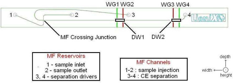
We present a simple approach in electrophoretic DNA separation and fluorescent monitoring that allows to identify the insertion or deletion of base-pairs in DNA probe molecules from genetic samples, and to perform intrinsic calibration/referencing for highly accurate DNA analysis. The principle is based on dual-point, dual-wavelength laser-induced fluorescence excitation using one or two excitation windows at the intersection of integrated waveguides and microfluidic channels in an optofluidic chip and a single, color-blind photodetector, resulting in a limit of detection of ~200 pM for single-end-labeled DNA molecules. The approach using a single excitation window is demonstrated experimentally, while the option exploiting two excitation windows is proposed theoretically.
Figures
Similar articles
-
Modulation-frequency encoded multi-color fluorescent DNA analysis in an optofluidic chip.Lab Chip. 2011 Feb 21;11(4):679-83. doi: 10.1039/c0lc00449a. Epub 2010 Dec 7. Lab Chip. 2011. PMID: 21140023
-
High-resolution electrophoretic separation and integrated-waveguide excitation of fluorescent DNA molecules in a lab on a chip.Electrophoresis. 2010 Aug;31(15):2584-8. doi: 10.1002/elps.201000126. Electrophoresis. 2010. PMID: 20665917
-
Fluorescence monitoring of microchip capillary electrophoresis separation with monolithically integrated waveguides.Opt Lett. 2008 Nov 1;33(21):2503-5. doi: 10.1364/ol.33.002503. Opt Lett. 2008. PMID: 18978901
-
Dual-color fluorescence cross-correlation spectroscopy on a planar optofluidic chip.Lab Chip. 2011 Apr 21;11(8):1502-6. doi: 10.1039/c0lc00401d. Epub 2011 Feb 22. Lab Chip. 2011. PMID: 21340094
-
[Laboratory on a microfluidic chip].Se Pu. 2005 Sep;23(5):456-63. Se Pu. 2005. PMID: 16350786 Review. Chinese.
Cited by
-
Optofluidic approaches for enhanced microsensor performances.Sensors (Basel). 2014 Dec 30;15(1):465-84. doi: 10.3390/s150100465. Sensors (Basel). 2014. PMID: 25558989 Free PMC article.
-
A hybrid silicon-PDMS optofluidic platform for sensing applications.Biomed Opt Express. 2014 Jan 9;5(2):417-26. doi: 10.1364/BOE.5.000417. eCollection 2014 Feb 1. Biomed Opt Express. 2014. PMID: 24575337 Free PMC article.
-
Frequency-encoded laser-induced fluorescence for multiplexed detection in infrared-mediated quantitative PCR.Analyst. 2014 Jun 7;139(11):2695-701. doi: 10.1039/c3an02334f. Analyst. 2014. PMID: 24448431 Free PMC article.
References
-
- Manz A., Graber N., Widmer H. M., “Miniaturized total chemical analysis systems: A novel concept for chemical sensing,” Sens. Actuators B Chem. 1, 244–248 (1990).
LinkOut - more resources
Full Text Sources
Miscellaneous