

Estimation of biological chromophores using diffuse optical spectroscopy: benefit of extending the UV-VIS wavelength range to include 1000 to 1600 nm
- PMID: 21258560
- PMCID: PMC3018130
- DOI: 10.1364/BOE.1.001432
Estimation of biological chromophores using diffuse optical spectroscopy: benefit of extending the UV-VIS wavelength range to include 1000 to 1600 nm
Abstract
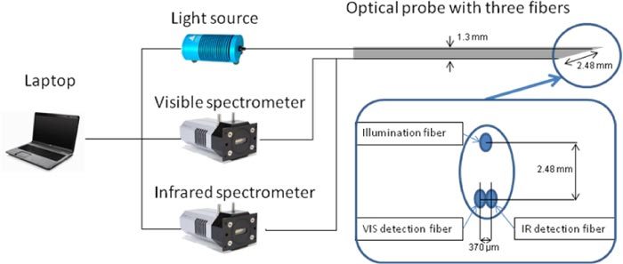
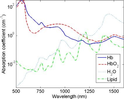
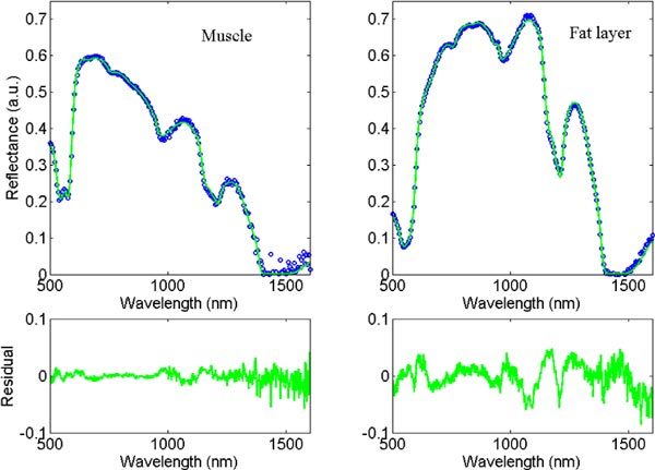
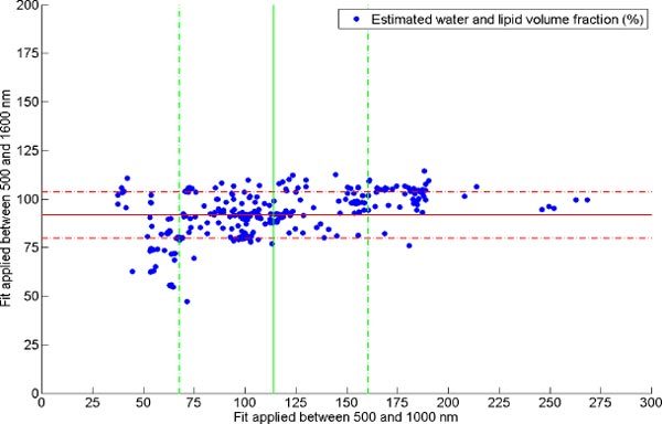
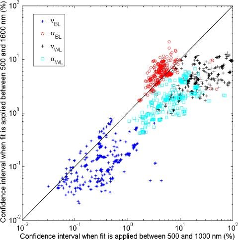
With an optical fiber probe, we acquired spectra from swine tissue between 500 and 1600 nm by combining a silicon and an InGaAs spectrometer. The concentrations of the biological chromophores were estimated by fitting a mathematical model derived from diffusion theory. The advantage of our technique relative to those presented in previous studies is that we extended the commonly-used wavelength ranges of 500 and 1000 nm to include the range of 1000 to 1600 nm, where additional water and lipid absorption features exist. Hence, a more accurate estimation of these two chromophores is expected when spectra are fitted between 500 and 1600 nm than between 500 and 1000 nm. When extending the UV-VIS wavelength range, the estimated total amount of chromophores approached 100% of the total as present in the probed volume. The confidence levels of the water and lipid related parameters increases by a factor of four.
Figures
Similar articles
-
Estimation of lipid and water concentrations in scattering media with diffuse optical spectroscopy from 900 to 1,600 nm.J Biomed Opt. 2010 May-Jun;15(3):037015. doi: 10.1117/1.3454392. J Biomed Opt. 2010. PMID: 20615044
-
Study of Minor Chromophores in Biological Tissues by Diffuse Optical Spectroscopy (Review).Sovrem Tekhnologii Med. 2025;17(1):146-162. doi: 10.17691/stm2025.17.1.12. Epub 2025 Feb 28. Sovrem Tekhnologii Med. 2025. PMID: 40071075 Free PMC article. Review.
-
InGaAs based gratings for UV-VIS spectrometer in prospective mRNA vaccine research.Opt Quantum Electron. 2022;54(9):555. doi: 10.1007/s11082-022-04002-1. Epub 2022 Jul 26. Opt Quantum Electron. 2022. PMID: 35912403 Free PMC article.
-
Effect of bile absorption coefficients on the estimation of liver tissue optical properties and related implications in discriminating healthy and tumorous samples.Biomed Opt Express. 2011 Feb 15;2(3):600-14. doi: 10.1364/BOE.2.000600. Biomed Opt Express. 2011. PMID: 21412465 Free PMC article.
-
UV-Vis Reflection-Absorption Spectroscopy at air-liquid interfaces.Adv Colloid Interface Sci. 2015 Nov;225:134-45. doi: 10.1016/j.cis.2015.08.012. Epub 2015 Sep 3. Adv Colloid Interface Sci. 2015. PMID: 26385430 Review.
Cited by
-
Review of short-wave infrared spectroscopy and imaging methods for biological tissue characterization.J Biomed Opt. 2015 Mar;20(3):030901. doi: 10.1117/1.JBO.20.3.030901. J Biomed Opt. 2015. PMID: 25803186 Free PMC article. Review.
-
Assessing spectral imaging of the human finger for detection of arthritis.Biomed Opt Express. 2019 Nov 26;10(12):6555-6568. doi: 10.1364/BOE.10.006555. eCollection 2019 Dec 1. Biomed Opt Express. 2019. PMID: 31853416 Free PMC article.
-
Diffuse reflectance spectroscopy of human liver tumor specimens - towards a tissue differentiating optical biopsy needle using light emitting diodes.Biomed Opt Express. 2018 Feb 8;9(3):1069-1081. doi: 10.1364/BOE.9.001069. eCollection 2018 Mar 1. Biomed Opt Express. 2018. PMID: 29541504 Free PMC article.
-
Epidural needle with embedded optical fibers for spectroscopic differentiation of tissue: ex vivo feasibility study.Biomed Opt Express. 2011 Jun 1;2(6):1452-61. doi: 10.1364/BOE.2.001452. Epub 2011 May 6. Biomed Opt Express. 2011. PMID: 21698009 Free PMC article.
-
Noninvasive assessment of hemodynamic and brain metabolism parameters following closed head injury in a mouse model by comparative diffuse optical reflectance approaches.Neurophotonics. 2016 Apr;3(2):025003. doi: 10.1117/1.NPh.3.2.025003. Epub 2016 May 9. Neurophotonics. 2016. PMID: 27175372 Free PMC article.
References
-
- Bard M. P., Amelink A., Hegt V. N., Graveland W. J., Sterenborg H. J. C. M., Hoogsteden H. C., Aerts J. G. J. V., “Measurement of hypoxia-related parameters in bronchial mucosa by use of optical spectroscopy,” Am. J. Respir. Crit. Care Med. 171(10), 1178–1184 (2005).10.1164/rccm.200501-046OC - DOI - PubMed
-
- Brown J. Q., Wilke L. G., Geradts J., Kennedy S. A., Palmer G. M., Ramanujam N., “Quantitative optical spectroscopy: a robust tool for direct measurement of breast cancer vascular oxygenation and total hemoglobin content in vivo,” Cancer Res. 69(7), 2919–2926 (2009).10.1158/0008-5472.CAN-08-3370 - DOI - PMC - PubMed
LinkOut - more resources
Full Text Sources
Other Literature Sources