. 2010 Oct 28;114(46):19569-19575.
doi: 10.1021/jp106453v.

Optical Properties of Single-Walled Carbon Nanotubes Separated in a Density Gradient; Length, Bundling, and Aromatic Stacking Effects

Affiliations

Optical Properties of Single-Walled Carbon Nanotubes Separated in a Density Gradient; Length, Bundling, and Aromatic Stacking Effects

Scott M Tabakman et al. J Phys Chem C Nanomater Interfaces. .

Abstract

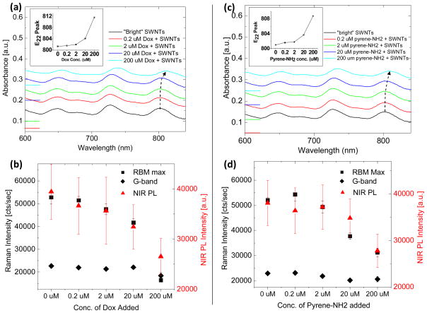
Single-walled carbon nanotubes (SWNTs) are promising materials for in vitro and in vivo biological applications due to their high surface area and inherent near infrared photoluminescence and Raman scattering properties. Here, we use density gradient centrifugation to separate SWNTs by length and degree of bundling. Following separation, we observe a peak in photoluminescence quantum yield (PL QY) and Raman scattering intensity where SWNT length is maximized and bundling is minimized. Individualized SWNTs are found to exhibit high PL QY and high resonance-enhanced Raman scattering intensity. Fractions containing long, individual SWNTs exhibit the highest PL QY and Raman scattering intensities, compared to fractions containing single, short SWNTs or SWNT bundles. Intensity gains of approximately ~1.7 and 4-fold, respectively, are obtained compared with the starting material. Spectroscopic analysis reveals that SWNT fractions at higher displacement contain increasing proportions of SWNT bundles, which causes reduced optical transition energies and broadening of absorption features in the UV-Vis-NIR spectra, and reduced PL QY and Raman scattering intensity. Finally, we adsorb small aromatic species on "bright," individualized SWNT sidewalls and compare the resulting absorption, PL and Raman scattering effects to that of SWNT bundles. We observe similar effects in both cases, suggesting aromatic stacking affects the optical properties of SWNTs in an analogous way to SWNT bundles, likely due to electronic structure perturbations, charge transfer, and dielectric screening effects, resulting in reduction of the excitonic optical transition energies and exciton lifetimes.


Figures

Figure 1
Figure 1
Centrifugation of sodium cholate-suspended single-walled carbon nanotubes (SWNTs) through a density gradient containing 1% sodium cholate, with discontinuous steps of 5%/10%/15%/20%/60% iodixanol at 50,000 RPM for one hour yielded a continuous distribution of SWNTs as well as a band formed at the 60% iodixanol boundary, as is clear from (a) photographs taken before and after DGC. Following aliquoting of 100 μL fractions (f#) as shown in (a) and normalization to the same optical density, photoluminescence under 808 nm excitation (b) showed varying quantum yields relative to the starting material (“start”), increasing from f3 to f6–7 and decreasing monotonically thereafter.
Figure 2
Figure 2
(a) 785 nm excitation Raman scattering spectra of DGC separated SWNTs at the same OD. Greater sensitivity is observed for the RBM peaks compared with the G-band peak, yet both features follow the same monotonic decrease in scattering intensity from f6-f23. An increase in intensity is observed for the RBM at 266 cm−1 corresponding to the (10,2) chirality (inset) with increasing fraction number. (b) Comparison of Raman scattering for the RBM at 233 cm−1 and G-band at 1590 cm−1 on the left axis with relative quantum yield, on the right axis, versus increasing fraction number. All three spectral features show an initial increase in intensity followed by a decrease. (c) Near-infrared absorption spectra for the DGC separated SWNT fractions shown in (a) as well as the cholate-SWNT starting material (dotted line). Red-shifting of the optical transition peaks was observed monotonically with increasing fraction number (curves are offset for clarity).
Figure 3
Figure 3
(a) Mean lengths and associated standard deviations of cholate-SWNTs measured by atomic force microscopy for DGC separated cholate-SWNTs fractions f4, f5, f6, f8, f14, and f20 in logarithmic scale. Lengths were calculated directly, without correction for tip convolution, for at least n=50 random SWNTs for fractions 4–14 and n=20 for f20. (b) Representative AFM images of SWNTs in fractions 4 and 6 to demonstrate the drastic increase in SWNT length.
Figure 4
Figure 4
Spectral properties of the SWNT optical transition near 800 nm. (a) Absorption peak position and full-width at half-maximum tend to increase with increasing fraction number. The arrows indicate the absorption peak position and FWHM, respectively, of the cholate-SWNT starting material, before DGC separation. (b) Peak intensity for the SWNT RBM feature at 266 cm−1, referred to as the “bundle peak,” following 785 nm excitation, with increasing fraction number. The solid lines represent moving averages (n=3) to aid in the observation of trends.
Figure 5
Figure 5
Non-covalent interaction of small aromatic molecules with DGC separated SWNTs causes similar effects as SWNT bundling. The loading of Doxorubicin (Dox) onto DGC separated, “bright” SWNTs causes a (a) concentration-dependent red-shifting of SWNT optical transition absorption peaks. Absorption spectra are offset vertically for clarity and zeroes are marked on the left axis. The peak position of the optical transition near 800 nm is plotted in the inset. (b) The intensity of the RBM at 233 cm−1 (black squares) as well as that of the G-band at 1590 cm−1 (black diamonds) decrease with increasing Dox concentration and subsequent optical transition red-shift. An accompanying decrease in photoluminescence intensity is also observed (red triangles). The loading of 1-pyrenemethylamine (pyrene-NH2) onto DGC-separated, “bright” SWNTs causes a similar, but slightly weaker effect, on (c) the SWNT optical transitions and (d) Raman scattering and photoluminescence intensities.

References

    1. O’Connell MJ, Bachilo SM, Huffman CB, Moore VC, Strano MS, Haroz EH, Rialon KL, Boul PJ, Noon WH, Kittrell C, Ma JP, Hauge RH, Weisman RB, Smalley RE. Band gap fluorescence from individual single-walled carbon nanotubes. Science. 2002;297:593–6. - PubMed
    1. Cherukuri P, Gannon CJ, Leeuw TK, Schmidt HK, Smalley RE, Curley SA, Weisman RB. Mammalian pharmacokinetics of carbon nanotubes using intrinsic near-infrared fluorescence. Proceedings of the National Academy of Sciences of the United States of America. 2006;103 (50):18882–18886. - PMC - PubMed
    1. Cherukuri P, Bachilo SM, Litovsky SH, Weisman RB. Near-infrared fluorescence microscopy of single-walled carbon nanotubes in phagocytic cells. Journal Of The American Chemical Society. 2004;126 (48):15638–15639. - PubMed
    1. Welsher K, Liu Z, Daranciang D, Dai H. Selective Probing and Imaging of Cells with Single Walled Carbon Nanotubes as Near-Infrared Fluorescent Molecules. Nano Letters. 2008;8 (2):586–590. - PubMed
    1. Leeuw TK, Reith RM, Simonette RA, Harden ME, Cherukuri P, Tsyboulski DA, Beckingham KM, Weisman RB. Single-walled carbon nanotubes in the intact organism: Near-IR imaging and biocompatibility studies in Drosophila. Nano Letters. 2007;7 (9):2650–2654. - PubMed

LinkOut - more resources