

Ethanol impairs glucose uptake by human astrocytes and neurons: protective effects of acetyl-L-carnitine
- PMID: 21258656
- PMCID: PMC3023411
Ethanol impairs glucose uptake by human astrocytes and neurons: protective effects of acetyl-L-carnitine
Abstract
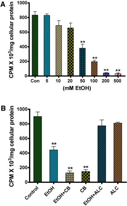
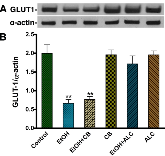
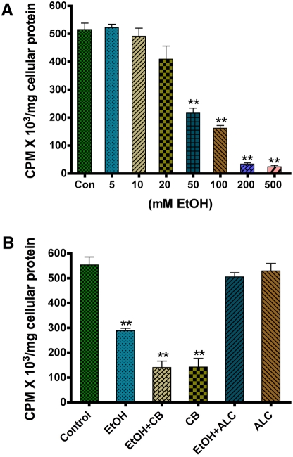
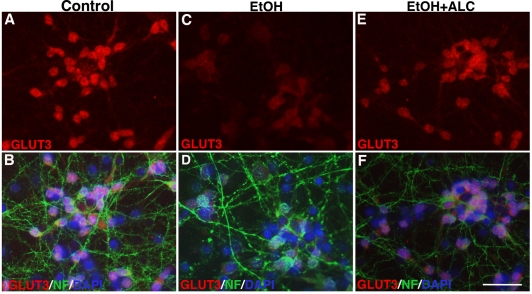
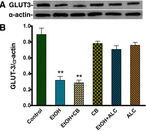
Alcohol consumption causes neurocognitive deficits, neuronal injury, and neurodegeneration. At the cellular level, alcohol abuse causes oxidative damage to mitochondria and cellular proteins and interlink with the progression of neuroinflammation and neurological disorders. We previously reported that alcohol inhibits glucose transport across the blood-brain barrier (BBB), leading to BBB dysfunction and neurodegeneration. In this study, we hypothesized that ethanol (EtOH)-mediated disruption in glucose uptake would deprive energy for human astrocytes and neurons inducing neurotoxicity and neuronal degeneration. EtOH may also have a direct effect on glucose uptake in neurons and astrocytes, which has not been previously described. Our results indicate that ethanol exposure decreases the uptake of D-(2-3H)-glucose by human astrocytes and neurons. Inhibition of glucose uptake correlates with a reduction in glucose transporter protein expression (GLUT1 in astrocytes and GLUT3 in neurons). Acetyl-L-carnitine (ALC), a neuroprotective agent, suppresses the effects of alcohol on glucose uptake and GLUT levels, thus reducing neurotoxicity and neuronal degeneration. These findings suggest that deprivation of glucose in brain cells contributes to neurotoxicity in alcohol abusers, and highlights ALC as a potential therapeutic agent to prevent the deleterious health conditions caused by alcohol abuse.
Keywords: Human astrocytes; acetyl-L-carnitine; glucose transporter protein; neurodegeneration.
IJPPP Copyright © 2011.
Figures

References
-
- WHO, editor. Geneva: WHO Statistical Information System (WHOSIS); 2007.
-
- Harper C. The neuropathology of alcohol-specific brain damage, or does alcohol damage the brain? J Neuropathol Exp Neurol. 1998;57:101–10. - PubMed
-
- Harper C, Dixon G, Sheedy D, Garrick T. Neuro-pathological alterations in alcoholic brains. Studies arising from the New South Wales Tissue Resource Centre. Prog Neuropsychopharmacol Biol Psychiatry. 2003;27:951–61. - PubMed
-
- Kurose I, Higuchi H, Kato S, Miura S, Watanabe N, Kamegaya Y, Tomita K, Takaishi M, Horie Y, Fukuda M, Mizukami K, Ishii H. Oxidative stress on mitochondria and cell membrane of cultured rat hepatocytes and perfused liver exposed to ethanol. Gastroenterology. 1997;112:1331–43. - PubMed
Grants and funding
LinkOut - more resources
Full Text Sources
Miscellaneous