

Review
doi: 10.1039/c0mt00061b.
Epub 2011 Jan 24.
Cupredoxins--a study of how proteins may evolve to use metals for bioenergetic processes
Affiliations
- PMID: 21258692
- PMCID: PMC6916721
- DOI: 10.1039/c0mt00061b
Item in Clipboard
Review
Cupredoxins--a study of how proteins may evolve to use metals for bioenergetic processes
Metallomics.
2011 Feb.
Abstract
Cupredoxins are small proteins that contain type I copper centers, which are ubiquitous in nature. They function as electron transfer shuttles between proteins. This review of the structure and properties of native cupredoxins, and those modified by site-directed mutagenesis, illustrates how these proteins may have evolved to specifically bind copper, develop recognition sites for specific redox partners, tune redox potential for a particular function, and allow for efficient electron transfer through the protein matrix. This is relevant to the general understanding of the roles of metals in energy metabolism, respiration and photosynthesis.
Figures
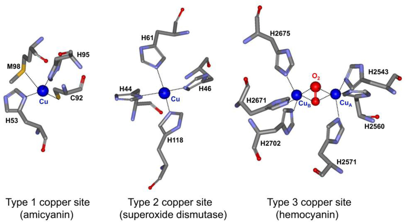
Examples of the three major classes of copper-binding sites in proteins. The structures were drawn using the structure coordinates from PDB files for the type 1 copper center of amicyanin from Paracoccus denitrificans (PDB code 2OV0), the type 2 copper center of Cu,Zn-superoxide dismutase from human bovine erythrocytes (PDB code 2SOD) and the type 3 copper center of hemocyanin from Octopus dofleini (PDB code 1JS8). The structures are presented as sticks colored gray for carbon, red for oxygen, blue for nitrogen and yellow for sulfur. The copper is displayed as a dark blue sphere.
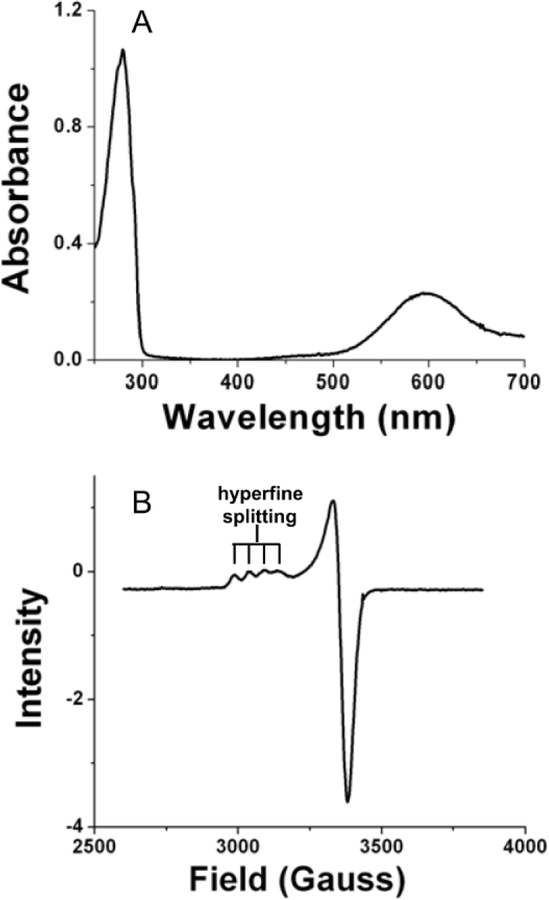
Spectroscopic properties of amicyanin which are characteristic of cupredoxins. (A) The visible absorption spectrum of oxidized (Cu2+) amicyanin. (B) X-band EPR spectrum of oxidized (Cu2+) amicyanin.
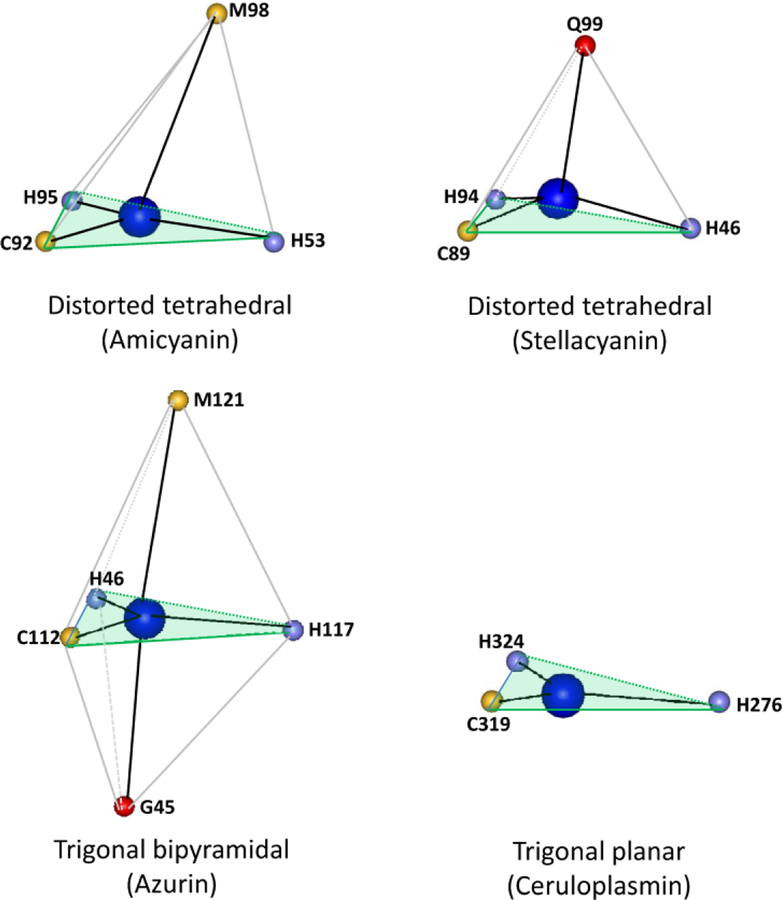
Variations in the ligation geometry of type 1 copper sites. The structures were drawn using the structure coordinates from PDB files for amicyanin from Paracoccus denitrificans (PDB code 2OV0), stellacyanin from Cucumis sativus (PDB code 1JER), azurin from Pseudomonas aeruginosa (PDB code 4AZU) and ceruloplasmin from human serum (PDB code 1KCW). Only the atom providing the copper ligand is shown with the one-letter amino acid code and residue number indicated. The plane described by the two His and one Cys ligand in each is shaded light green.

The methylamine dehydrogenase-amicyanin-cytochrome c-551i complex. One half of the symmetrical complex of the crystal structure (PDB code 2MTA) is shown with the cytochrome colored tan, amicyanin colored blue, the MADH α subunit colored olive green, and the MADH β subunit colored light green. The TTQ in MADH, copper in amicyanin and heme in the cytochrome are colored black.

Structures of representative cupredoxins. The secondary structures are highlighted with beta sheets colored light blue, alpha helices colored red, and unstructured loops and turns colored gray. The structures were drawn using the structure coordinates from PDB files for amicyanin from Paracoccus denitrificans (PDB code 2OV0), plastocyanin from Ulva pertusa (PDB code 1IUZ), azurin from Pseudomonas aeruginosa (PDB code 4AZU) and stellacyanin from Cucumis sativus (PDB code 1JER).
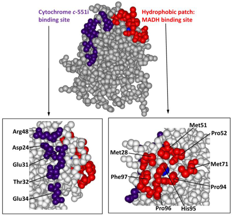
Space filling model of the structure of amicyanin from Paracoccus denitrificans (PDB code 2OV0). The MADH binding site (red) seen in the structure of the protein complex is comprised of residues Met28, Met51, Pro52, Met71, Pro94, Pro96 and Phe97. The cytochrome c-551i binding site (purple) is comprised of Asp24, Glu31, Glu32, Thr34 and Arg48. The copper is displayed as a blue sphere.

Redox partner binding sites of plastocyanins. Space filling models of the structure were prepared using the coordinates from the PDB files of plastocyanins from a higher plant (Silene PDB code 1BYO), from a green alga (Ulva pertusa PDB code 1IUZ) and from a cyanobacteria (Synechococuss sp. PDB code 1BXU). The hydrophobic patch which is the site of interaction with cytochrome f and is present in all three is red. The acidic patch which also interacts with cytochrome f in the plastocyanins of plants and algae is purple. The additional acid patch found only in plastocyanin from plants is green. Copper is indicated as a blue sphere.
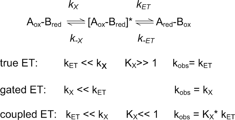
Kinetic mechanisms of electron transfer reactions
References
Publication types
MeSH terms
Substances
Grants and funding
LinkOut - more resources
Full Text Sources
