

Membrane localization of insulin receptor substrate-2 (IRS-2) is associated with decreased overall survival in breast cancer
- PMID: 21258861
- PMCID: PMC3128655
- DOI: 10.1007/s10549-011-1353-1
Membrane localization of insulin receptor substrate-2 (IRS-2) is associated with decreased overall survival in breast cancer
Abstract
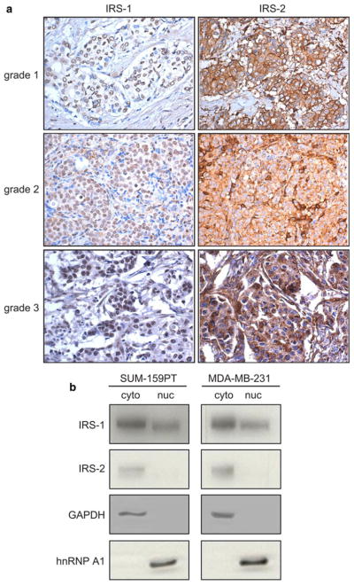
Recent studies have identified a role for insulin receptor substrate-2 (IRS-2) in promoting motility and metastasis in breast cancer. However, no published studies to date have examined IRS-2 expression in human breast tumors. We examined IRS-2 expression by immunohistochemistry (IHC) in normal breast tissue, benign breast lesions, and malignant breast tumors from the institutional pathology archives and a tumor microarray from a separate institution. Three distinct IRS-2 staining patterns were noted: diffusely cytoplasmic, punctate cytoplasmic, and localized to the cell membrane. The individual and pooled datasets were analyzed for associations of IRS-2 staining pattern with core clinical parameters and clinical outcomes. Univariate analysis revealed a trend toward decreased overall survival (OS) with IRS-2 membrane staining, and this association became significant upon multivariate analysis (P = 0.01). In progesterone receptor negative (PR-) tumors, in particular, IRS-2 staining at the membrane correlated with significantly worse OS than other IRS-2 staining patterns (P < 0.001). When PR status and IRS-2 staining pattern were evaluated in combination, PR- tumors with IRS-2 at the membrane were associated with a significantly decreased OS when compared with all other combinations (P = 0.002). Evaluation of IRS-2 staining patterns could potentially be used to identify patients with PR- tumors who would most benefit from aggressive treatment.
Figures


References
-
- Furlanetto RW, DiCarlo JN. Somatomedin-C receptors and growth effects in human breast cells maintained in long-term tissue culture. Cancer Res. 1984;44(5):2122–2128. - PubMed
-
- Turner BC, Haffty BG, Narayanan L, Yuan J, Havre PA, Gumbs AA, Kaplan L, Burgaud JL, Carter D, Baserga R, Glazer PM. Insulin-like growth factor-I receptor overexpression mediates cellular radioresistance and local breast cancer recurrence after lumpectomy and radiation. Cancer Res. 1997;57(15):3079–3083. - PubMed
-
- Johnston SR, Dowsett M, Smith IE. Towards a molecular basis for tamoxifen resistance in breast cancer. Ann Oncol. 1992;3(7):503–511. - PubMed
-
- Resnik JL, Reichart DB, Huey K, Webster NJ, Seely BL. Elevated insulin-like growth factor I receptor autophosphorylation and kinase activity in human breast cancer. Cancer Res. 1998;58(6):1159–1164. - PubMed
-
- White MF, Maron R, Kahn CR. Insulin rapidly stimulates tyrosine phosphorylation of a Mr-185, 000 protein in intact cells. Nature. 1985;318(6042):183–186. - PubMed
Publication types
MeSH terms
Substances
Grants and funding
LinkOut - more resources
Full Text Sources
Other Literature Sources
Medical
Research Materials
