. 2011 Mar 15;50(10):1651-63.
doi: 10.1021/bi101736v. Epub 2011 Feb 15.

Intermonomer electron transfer between the low-potential b hemes of cytochrome bc₁

Affiliations

Intermonomer electron transfer between the low-potential b hemes of cytochrome bc₁

Pascal Lanciano et al. Biochemistry. .

Abstract

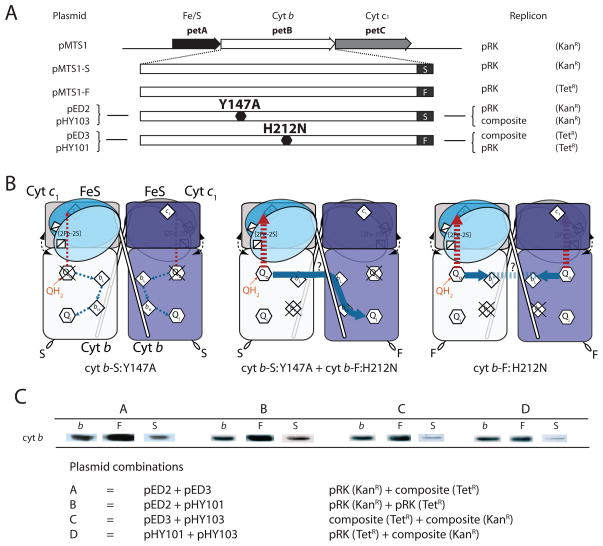
Cytochrome (cyt) bc(1) is a structural dimer with its monomers consisting of the Fe-S protein, cyt b, and cyt c(1) subunits. Its three-dimensional architecture depicts it as a symmetrical homodimer, but the mobility of the head domain of the Fe-S protein indicates that the functional enzyme exists in asymmetrical heterodimeric conformations. Here, we report a new genetic system for studying intra- and intermonomer interactions within the cyt bc(1) using the facultative phototrophic bacterium Rhodobacter capsulatus. The system involves two different sets of independently expressed cyt bc(1) structural genes carried by two plasmids that are coharbored by a cell without its endogenous enzyme. Our results indicate that coexpressed cyt bc(1) subunits were matured, assorted, and assembled in vivo into homo- and heterodimeric enzymes that can bear different mutations in each monomer. Using the system, the occurrence of intermonomer electron transfer between the low-potential b hemes of cyt bc(1) was probed by choosing mutations that perturb electron transfer at the hydroquinone oxidation (Q(o)) and quinone reduction (Q(i)) sites of the enzyme. The data demonstrate that active heterodimeric variants, formed of monomers carrying mutations that abolish only one of the two (Q(o) or Q(i)) active sites of each monomer, are produced, and they support photosynthetic growth of R. capsulatus. Detailed analyses of the physicochemical properties of membranes of these mutants, as well as purified homo- and heterodimeric cyt bc(1) preparations, demonstrated that efficient and productive electron transfer occurs between the low-potential b(L) hemes of the monomers in a heterodimeric enzyme. Overall findings are discussed with respect to intra- and intermonomer interactions that take place during the catalytic turnover of cyt bc(1).


Figures

Figure 1
Figure 1. The three dimensional structure of cyt bc1 dimeric and its cofactors
The three dimensional structure (PDB # 1ZRT for R. capsulatus) (left) of cyt bc1 depicts it as a dimeric enzyme with two identical monomers, M1 and M2, each formed of the Fe/S protein (light blue), cyt b (dark blue in M1 and yellow in M2) and cyt c1 (beige). The figure is generated using Pymol (DeLano Scientific, San Carlos, CA), and the cofactors of each monomer ([2Fe2S] cluster, hemes bL and bH and c1) are shown in different colors (blue in M1 and red in M2). Black and red arrows indicate the electron transfer pathways between the cofactors and the Qo/Qi active sites of cyt bc1, respectively. Inter-monomer hemes bLbL distance is indicated with a dotted line, but for the sake of clarity the distances (11) separating the cofactors are not shown. The right panel shows a schematic depiction of a homo-dimeric cyt bc1, with its cofactors and Qo/Qi active sites, as described for the left panel.
Figure 2
Figure 2. A genetic system to study intra- and inter-monomer interactions in cyt bc1 dimer: cyt b-S:Y147A + cyt b-F:H212N hetero-dimeric cyt bc1 variant in R. capsulatus
A. The petABC of R. capsulatus is composed of petA, petB and petC genes encoding the Fe/S protein, cyt b and cyt c1 subunits, respectively. The different plasmids carrying this operon (left), their replicon type and antibiotic resistance markers (right), as well as the location of cyt b mutations in petB (•, middle) that they carry are shown together with the Strep (S) and Flag (F) epitope tags added at the carboxyl terminal end of cyt b. The brackets are to indicate that pED2 and pHY103 carry the same cyt b mutation but in two different replicons with the same antibotic resistance marker, and the same is also the case for pED3 and pHY101. B. Co-expression of a KanR plasmid carrying petABC with cyt b-S:Y147A mutation and a TetR plasmid carrying petABC with cyt b-F:H212N mutation yields the homo-dimeric cyt b-S:Y147A (left) and cyt b-F:H212N (right) as well as the hetero-dimeric cyt b-S:Y147A + cyt b-F:H212N (middle) cyt bc1 variants. The thin dotted arrows refer to residual electron transfer that occurs in the homo-dimeric cyt b-S:Y147A variant, whereas the thick red dashed arrows, and thick blue solid arrows correspond to electron transfer to the high and low potentials cofactors of cyt bc1, respectively. Note that inter-monomer hemes bL to bL electron transfer is indicated by a thick blue solid arrow. C. SDS/PAGE-immunoblot analyses of Strep- and Flag-tagged cyt b in chromatophore membranes of strains harboring different combinations of KanR and TetR plasmids (A to D, as indicated) producing hetero-dimeric cyt bc1 variants. Approximately 10 μg of total proteins were used and cyt b contents were determined using polyclonal antibodies specific to R. capsulatus cyt b (b) as well as commercially available antibodies against the Strep (S) and Flag (F) epitope tags. Properties of the plasmids used are described in Table 1.
Figure 3
Figure 3. Photosynthetic growth phenotypes and biochemical properties of R. capsulatus strains producing hetero-dimeric cyt bc1 variants
A. Photosynthetic (Ps) growth on enriched medium of R. capsulatus strains harboring plasmids pMTS1, pMTS1-S and pMTS1-F producing wild type, pED2 and pED3 producing homo-dimeric cyt b-S:Y147A and cyt b-F:H212N, and pED2+pED3 producing hetero-dimeric cyt b-S:Y147A + cyt b-F:H212N cyt bc1 variants are shown. Cells harboring pMTS1, pMTS1-S and pMTS1-F are Ps+, those harboring pED2 or pED3 are Ps, and those harboring both pED2 and pED3 are Ps+/− (i. e., slow growth). Shown above the plate a small section where rare Ps+ revertants in pED2+pED3 cultures (indicated by arrows) are observed. B. Chromatophore membranes of appropriate R. capsulatus strains described in A and grown by respiration in enriched medium were analyzed by SDS-PAGE (15%)/immunoblots using approximately 10 μg of total proteins. The cyt b and cyt c1 subunits were determined using polyclonal anti-cyt b or anti-cyt c1 (right) or anti-Strep II and anti-Flag M2 (left) antibodies as indicated. C. Total b-type and c-type cyts contents of chromatophore membranes (0.3 mg/mL) obtained from appropriate R. capsulatus strains (as described in A) grown by respiration in enriched medium were determined by ascorbate (dashed lines) or dithionite (solid lines) reduced minus ferricyanide oxidized optical difference spectra. Note the differences seen between the stains harboring different plasmids and producing different cyt bc1 variants. D. Low spin heme EPR spectra from redox-poised chromatophore membranes obtained from appropriate R. capsulatus strains (as described in A) grown by respiration in enriched medium. The ambient redox potential (Eh) values at which the spectra were recorded were 255, 260, 300 and 290 mV for membranes derived from cells harboring pMTS1, pED2, pED3 and pED2+pED3 plasmids, respectively. Experimental conditions used were as follows: temperature, 10K; microwave power, 10 mW at 9.420 GHz; modulation amplitude, 10 G at 100 kHz, number of scan, 4, and note that no baseline correction was used. E. [2Fe-2S] EPR spectra of redox-poised chromatophore membranes obtained from appropriate R. capsulatus strains (as described in A) grown by respiration in enriched medium. The Eh values at which the spectra were recorded were 123, 112, 116 and 126 mV for membranes derived from cells harboring pMTS1, pED2, pED3 and pED2+pED3 plasmids, respectively. Experimental conditions used were as follows: temperature, 20K; microwave power, 2 mW at 9.416 GHz; modulation amplitude, 20 G at 100 kHz; number of scan, 1.
Figure 4
Figure 4. Biochemical properties of partially purified R. capsulatus cyt b-S:Y147A + cyt b-F:H212N hetero-dimeric cyt bc1 variants
A. Homo-dimeric (FF and SS) and hetero-dimeric (FS) cyts bc1 variants purified from cells harboring pED2+pED3 by DEAE-Biogel chromatography were analyzed by SDS-PAGE (12.5%)/immunoblots using approximately 5 μg proteins and anti-Strep II (α-Strep) or anti-Flag M2 (α-Flag) monoclonal antibodies. Fe/S protein, cyt b and cyt c1 subunits of cyt bc1 are indicated. Note that Flag tagged (pMTS1-F) and Strep tagged (pMTS1-F) wild type, as well as homodimeric cyt b-S:Y147A (pED2) and cyt b-F:H212N (pED3) cyt bc1 variants were also purified, but not shown for the sake of clarity. B. Total b-type and c-type cyts contents of purified materials obtained from appropriate R. capsulatus strains (as described in Fig. 2A) were used to determine their ascorbate (dashed lines) or dithionite (solid lines) reduced minus ferricyanide oxidized optical difference spectra. Note that thee spectra are similar to those obtained using appropriate chromatophore membranes shown in Fig 3C. C. Low spin heme EPR spectra of purified materials obtained from appropriate R. capsulatus strains (as described in Fig. 2A). All samples were oxidized directly in the EPR tube by adding 5μL 10mM ferricyanide. The experimental conditions were as in Fig. 3D, and a “non signal” portion of a baseline spectrum simulated by a third-order polynomial fit subtracted as baseline correction.
Figure 5
Figure 5. Light-induced, time-resolved cyt b reduction and cyt c re-reduction kinetics of various R. capsulatus strains
R. capsulatus strains harboring plasmids pMTS1 producing wild type, pED2 and pED3 producing homo-dimeric cyt b-S:Y147A and cyt b-F:H212N, respectively, and pED2+pED3 producing hetero-dimeric cyt b-S:Y147A + cyt b-F:H212N cyt bc1 variants were analyzed. In each case, chromatophore membranes corresponding to an amount of RC equal to 0.30 μM were resuspended in 50 mM MOPS buffer (pH 7.0) containing 100 mM KCl at an Eh of 100 mV. The amount of RC in each case was determined from the extent of its photo-oxidation after a train of 10 flashes, separated by 50 ms at an Eh of 380 mV (upper panel) and an extinction coefficient of 29 mM−1 cm−1 at 605 minus 540 nm, as described in Material and Methods. In all samples, cyt c re-reduction kinetics (middle panel) were monitored at 550 minus 540 nm, in the absence of inhibitor (no), or in the presence of 10 μM myxothiazol (Myx) which abolishes QH2 oxidation at the Qo site, or 10 μM stigmatellin (Stig), which abolishes electron transfer from the [2Fe-2S] cluster to heme c1 by immobilizing the head domain of the Fe/S protein subunit at the Qo site. Similarly, cyt b reduction kinetics (lower panel) were monitored at 560 minus 570 nm, in the absence of inhibitor (no), or in the presence of 10 μM of the Qi site inhibitor antimycin (Ant). Note that the samples of pED2 were analyzed using slightly different equipment, which yielded a higher signal to noise ratio than the other samples.
Figure 6
Figure 6. Light-induced, time-resolved cyt b reduction kinetics exhibited by R. capsulatus strains harboring different plasmid combinations yielding hetero-dimeric cyt bc1 variants
The cyt b reduction kinetics were monitored using chromatophore membranes prepared from appropriate cells (pMTS1, pED2+pED3, pED3+pHY103, pHY101+pHY103 and pED2+pED3) grown in enriched medium either under respiratory growth conditions. All plasmid combinations (A to D) are described in Fig. 2 and Table 1. Experimental conditions were as described in Fig. 5, using chromatophore membranes containing 0.30 μM of RC, appropriate mediators (Materials and Methods), 3 μM valinomycin, and 10 μM antimycin A. Samples were poised at 100 mV. Note that the extent and rate of cyt b reduction changes depending on the strains, with cells harboring identical replicons exhibiting more pronounced kinetics, possibly reflecting better electronic couplings in membranes harboring heterogenous cyt bc1 subpopulations.
Figure 7
Figure 7. A model depicting inter-monomer electron transfer between the hemes bL of monomers in a hetero-dimeric cyt bc1
The three dimensional structure (1ZRT) of R. capsulatus cyt bc1 shown in Fig. 1 is modified to show (front view, left and top view, right) the location and nature of the mutations used in this work, revealing inter-monomer electron transfer between the hemes bL cofactors of the enzyme. All structural features are as in Fig. 1, except that the cyt b:Y147A and cyt b:H212N substitutions are shown as space filling models in blue on M1 and red on M2 monomers. The absence of heme bH in one monomer, due to the latter mutation, is indicated by a black ellipsoid, and the residual electron transfer that is unable to support Ps growth but still occurs in the presence of the former mutation is indicated by dotted arrows. The thick black arrows correspond to the productive inter-monomer electron transfer that occurs across the functional Qo and Qi sites of the hetero-dimeric cyt bc1. Note that this electron transfer is efficient enough to support Ps growth of R. capsulatus but apparently is rate limiting because it can only confer a slow Ps+/− growth phenotype.

References

    1. Berry EA, Guergova-Kuras M, Huang LS, Crofts AR. Structure and function of cytochrome bc complexes. Annu Rev Biochem. 2000;69:1005–1075. - PubMed
    1. Mitchell P. Possible molecular mechanisms of the protonmotive function of cytochrome systems. J Theor Biol. 1976;62:327–367. - PubMed
    1. Trumpower BL. The protonmotive Q cycle. Energy transduction by coupling of proton translocation to electron transfer by the cytochrome bc1 complex. J Biol Chem. 1990;265:11409–11412. - PubMed
    1. Daldal F, Davidson E, Cheng S. Isolation of the structural genes for the Rieske Fe-S protein, cytochrome b and cytochrome c1 all components of the ubiquinol: cytochrome c2 oxidoreductase complex of Rhodopseudomonas capsulata. J Mol Biol. 1987;195:1–12. - PubMed
    1. Atta-Asafo-Adjei E, Daldal F. Size of the amino acid side chain at position 158 of cytochrome b is critical for an active cytochrome bc1 complex and for photosynthetic growth of Rhodobacter capsulatus. Proc Natl Acad Sci U S A. 1991;88:492–496. - PMC - PubMed

Publication types

MeSH terms