. 2008 May;1(3):247-57.
doi: 10.1111/j.1751-7915.2008.00028.x.

PEGylation of bacteriophages increases blood circulation time and reduces T-helper type 1 immune response

Affiliations

PEGylation of bacteriophages increases blood circulation time and reduces T-helper type 1 immune response

Kwang-Pyo Kim et al. Microb Biotechnol. 2008 May.

Abstract

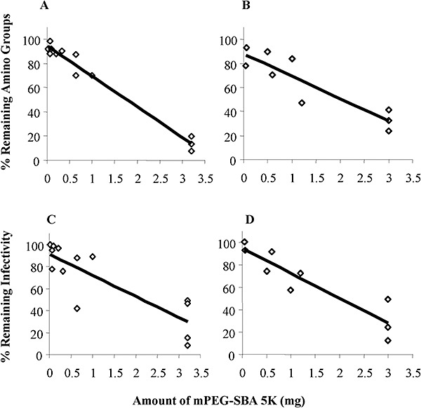
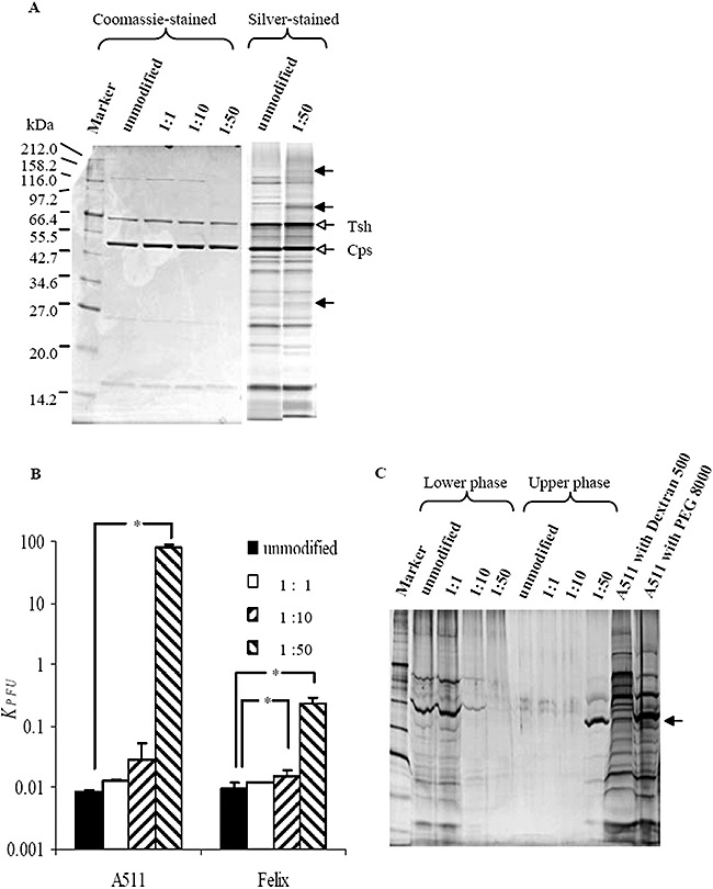
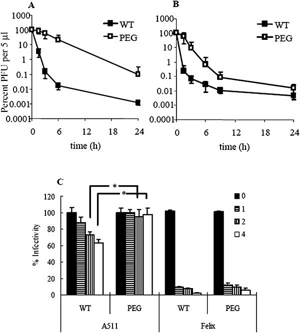
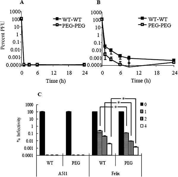
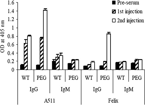
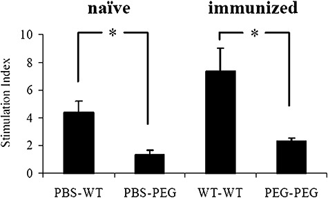
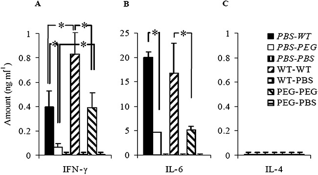
The increasing occurrence of antibiotic-resistant pathogens is of growing concern, and must be counteracted by alternative antimicrobial treatments. Bacteriophages represent the natural enemies of bacteria. However, the strong immune response following application of phages and rapid clearance from the blood stream are hurdles which need to be overcome. Towards our goal to render phages less immunogenic and prolong blood circulation time, we have chemically modified intact bacteriophages by conjugation of the non-immunogenic polymer monomethoxy-polyethylene glycol (mPEG) to virus proteins. As a proof of concept, we have used two different polyvalent and strictly virulent phages of the Myoviridae, representing typical candidates for therapeutical approaches: Felix-O1 (infects Salmonella) and A511 (infects Listeria). Loss of phage infectivity after PEGylation was found to be proportional to the degree of modification, and could be conveniently controlled by adjusting the PEG concentration. When injected into naïve mice, PEGylated phages showed a strong increase in circulation half-life, whereas challenge of immunized mice did not reveal a significant difference. Our results suggest that the prolonged half-life is due to decreased susceptibility to innate immunity as well as avoidance of cellular defence mechanisms. PEGylated viruses elicited significantly reduced levels of T-helper type 1-associated cytokine release (IFN-γ and IL-6), in both naïve and immunized mice. This is the first study demonstrating that PEGylation can increases survival of infective phage by delaying immune responses, and indicates that this approach can increase efficacy of bacteriophage therapy.


Figures

Figure 1
Figure 1
Effect of PEGylation on infectivity of phage. Determination of the degree of PEGylation by measuring the percentage of remaining unmodified amino groups (A, A511; B, Felix‐O1), and the percentage of infective particles after PEGylation (C, A511; D, Felix‐O1). Trend lines are shown.
Figure 2
Figure 2
A. An SDS‐PAGE analysis of unmodified and PEGylated A511, stained with Coomassie or Silver. The major structural proteins (capsid Cps and tail Tsh) are marked by open arrows, and possible protein modifications/gel shifts are indicated by solid arrows.
B and C. Results of the partitioning assay (see text for details). The ratio of infective particles in the upper and lower phases is shown for both A511 and Felix‐O1 (B). The asterisk (*) indicates statistical significance (α = 0.05). (C) shows an SDS‐PAGE analysis of phage A511 native and PEGylated proteins after recovery from the partitioning assay (see text). The phase transition of Cps (indicated by an arrow) is clearly evident. Samples of 30 µl were loaded and silver‐stained after electrophoresis. As controls, native A511 mixed with either Dextran 500 or PEG 8000 was also analysed.
Figure 3
Figure 3
Phage inactivation in the presence of innate immunity in the mouse model, both in vivo for A511 (A) and Felix‐O1 (B), and in vitro (C). In (C), 0, 1, 2 and 4 indicate the incubation time in the presence of serum. In in vivo studies, the number of phage per 5 µl at 0 h is represented as 100%, corresponded to 1 × 106 PFU for WT A511 and 1 × 105 PFU for PEGylated A511, and 1 × 106 PFU for WT Felix‐O1 and 3 × 105 PFU for PEGylated Felix‐O1. The asterisk (*) denotes statistical significance (α = 0.05).
Figure 4
Figure 4
Phage inactivation in vivo for A511 (A) and Felix‐O1 (B), or in vitro (C) in the presence of adaptive immunity. In in vivo studies, the number of phage per 5 µl at 0 h is represented as 100%, corresponded to 1 × 106 PFU for WT A511 and 1 × 105 PFU for PEGylated A511, and 1 × 106 PFU for WT Felix‐O1 and 3 × 105 PFU for PEGylated Felix‐O1. In (C), 0, 1, 2 and 4 indicates the incubation time in the presence of serum. The asterisk (*) denotes statistical significance (α = 0.05).
Figure 5
Figure 5
Amounts of antigen‐specific IgG and IgM in pre‐serum, serum taken 12 days after the first injection and serum taken 7 days after the second immunization. The antigens used for ELISA plate coating and immunization were identical.
Figure 6
Figure 6
Stimulation of splenocyte proliferation by bacteriophage Felix‐O1. Mice splenocytes were used, which were prepared from either naïve mice (left bars) or immunized animals which have received two doses from the same antigen (right bars) either native (WT) or PEGylated Felix‐O1 (PEG). Phosphate‐buffered saline (PBS) was used as a control. The asterisk (*) indicates statistical significance (α = 0.05).
Figure 7
Figure 7
Cytokine induction by unmodified and PEGylated Felix‐O1 phage, in naïve (italics) and in immunized mice. Splenocytes from naïve or immunized mice which were consecutively treated with the same antigens were used. The legend indicates the immunizing antigens and the treatment antigens, either unmodified phage (WT), PEGylated phage (PEG) or buffer (PBS). (A) IFN‐γ; (B) IL‐6; (C) IL‐4. The asterisk (*) denotes statistically significant differences (α = 0.05).

Similar articles

Cited by

References

    1. Abuchowski A., McCoy J.R., Palczuk N.C., Van Es T., Davis F.F. Effect of covalent attachment of polyethylene glycol on immunogenicity and circulating life of bovine liver catalase. J Biol Chem. 1977a;11:3582–3586. - PubMed
    1. Abuchowski A., Van Es T., Palczuk N.C., Davis F.F. Alteration of immunological properties of bovine serum albumin by covalent attachment of polyethylene glycol. J Biol Chem. 1977b;11:3578–3581. - PubMed
    1. Ackermann H.W., Nguyen T.M. Sewage coliphages studied by electron microscopy. Appl Environ Microbiol. 1983;45:1049–1059. - PMC - PubMed
    1. Adams M.H. Methods of study of bacterial viruses. In: Hershey A.D., editor. Interscience Publishers; 1959. pp. 443–457.
    1. Alemany R., Suzuki K., Curiel D.T. Blood clearance rates of adenovirus type 5 in mice. J Gen Virol. 2000;81:2605–2609. - PubMed

Publication types

MeSH terms

Substances