. 2009 May;2(3):343-60.
doi: 10.1111/j.1751-7915.2009.00093.x. Epub 2009 Feb 23.

A molecular key for building hyphae aggregates: the role of the newly identified Streptomyces protein HyaS

Affiliations

A molecular key for building hyphae aggregates: the role of the newly identified Streptomyces protein HyaS

Ilona Koebsch et al. Microb Biotechnol. 2009 May.

Abstract

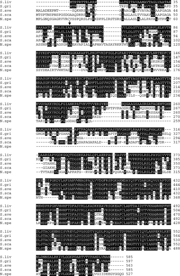
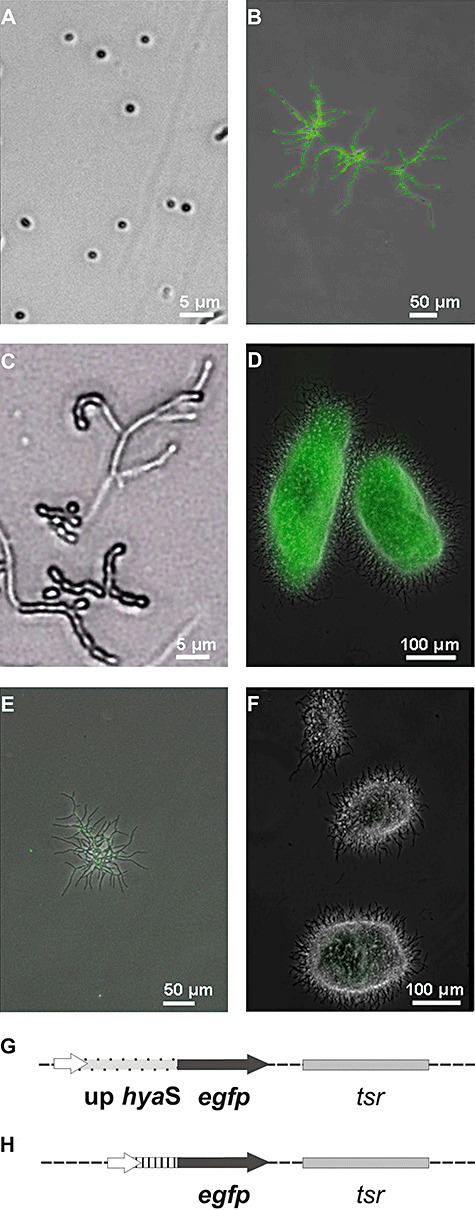
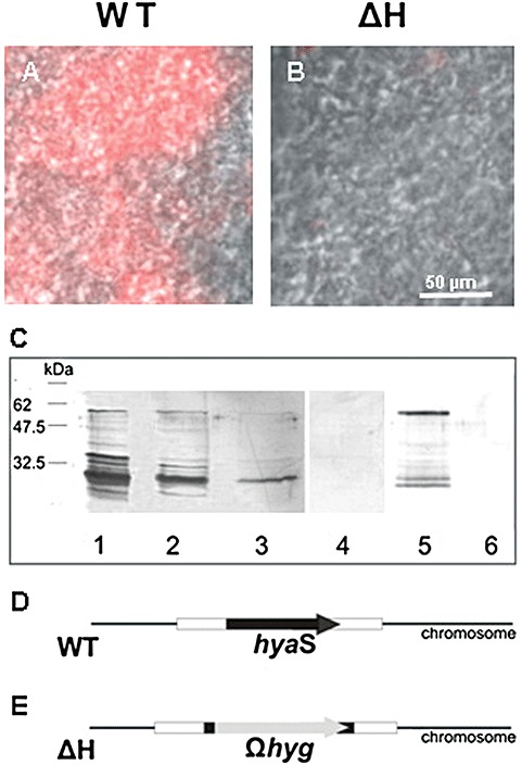
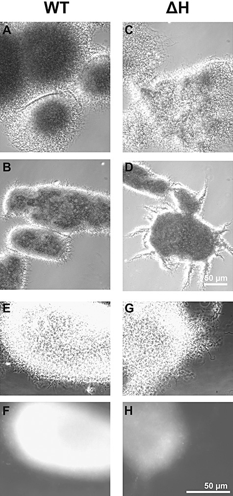
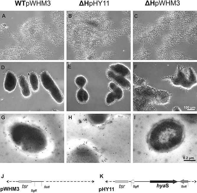
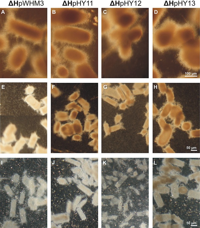
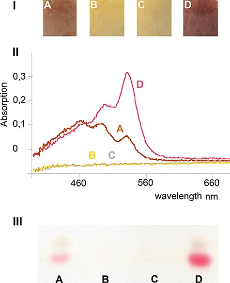
Streptomycetes produce many metabolites with medical and biotechnological applications. During fermentations, their hyphae build aggregates, a process in which the newly identified protein HyaS plays an important role. The corresponding hyaS gene is present within all investigated Streptomyces species. Reporter fusions indicate that transcription of hyaS occurs within substrate hyphae of the Streptomyces lividans wild type (WT). The HyaS protein is dominantly associated with the substrate hyphae. The WT strain forms cylindrically shaped clumps of densely packed substrate hyphae, often fusing to higher aggregates (pellets), which remain stably associated during shaking. Investigations by electron microscopy suggest that HyaS induces tight fusion-like contacts among substrate hyphae. In contrast, the pellets of the designed hyaS disruption mutant ΔH are irregular in shape, contain frequently outgrowing bunches of hyphae, and fuse less frequently. ΔH complemented with a plasmid carrying hyaS resembles the WT phenotype. Biochemical studies indicate that the C-terminal region of HyaS has amine oxidase activity. Investigations of ΔH transformants, each carrying a specifically mutated gene, lead to the conclusion that the in situ oxidase activity correlates with the pellet-inducing role of HyaS, and depends on the presence of certain histidine residues. Furthermore, the level of undecylprodigiosin, a red pigment with antibiotic activity, is influenced by the engineered hyaS subtype within a strain. These data present the first molecular basis for future manipulation of pellets, and concomitant production of secondary metabolites during biotechnological processes.


Figures

Figure 1
Figure 1
Alignment of HyaS with other hypothetical proteins. Block comparisons of deduced gene products from the streptomycetes S. lividans (S.liv, EMBL, DS: 72304), S. griseus (S.gri, SGR_3840), S. avermitilis (S.ave, SAV_4459), S. scabies (S.sca, 4945557‐4943764), Nocardioides sp. JS614 (N.spe, Noca_1817). The predicted S. coelicolor A3(2) protein (SCO 7657) corresponded to 99% to that of S. lividans; hence, it is not aligned. Amino acid residues, which are identical with the S. lividans protein, are marked white on a black background. The cleavage site, to generate the signal peptide, is indicated by an arrow.
Figure 2
Figure 2
Synthesis of EGFP.
A–D. The S. lividans transformant containing the construct (G) was analysed as spores (A), during early growth – as young substrate hyphae – on solid medium (B), during formation of aerial hyphae (C), or after cultivation in a flask filled with liquid medium under shaking conditions for 20 h (D).
E and F. The S. lividans transformant with the control plasmid (H) was analysed (E and F) under the conditions presented under (B) or (D). Pictures were taken under visual light and under UV light using a Zeiss Axiovert microscope; merged pictures are presented (A–F). The magnification is given for each picture as a bar.
G. The plasmid construct is a pWHM3 derivative, containing the egfp gene (black arrow) in frame with the upstream region of the hyaS gene (light grey with black points).
H. The control plasmid corresponds to that one presented under (G), except that it has a small DNA fragment lacking the regulatory region (stripes) in front of egfp.
Figure 3
Figure 3
Localization of the HyaS protein, relevant characteristics of the chromosome from S. lividans WT and ΔH, and plasmid constructs.
A and B. The hyphae of S. lividans WT (A) or ΔH (B) were grown in complete liquid medium without shaking for 17 h. Aliquots (A or B) were placed onto a polylysine‐coated glass slide (see Experimental procedures), treated with primary anti‐HyaS antibodies, and with Alexa Fluor 647‐coupled secondary rat antibodies. Subsequently, slides were analysed under UV light with a filter set for Cy5 and by phase‐contrast microscopy. The resulting pictures were merged; the bar in (B) indicates the magnification for (A) and (B).
C. WT mycelia – grown as described above – were collected by centrifugation, washed successively three times with 1 M NaCl, and aliquots (lanes 1–3) were separated by SDS‐PAGE. As control, ΔH mycelia grew in the same fashion, and washings with 1 M NaCl (lane 4, aliquot of the first wash) were analysed. The proteins of the supernatant were precipitated by ammonium sulfate (90% w/v), re‐suspended and each sample (WT, lane 5 and ΔH, lane 6) was separated by SDS‐PAGE, and then transferred to a nylon membrane. This was treated with primary anti‐HyaA antibodies, then with secondary anti‐rat antibodies conjugated with alkaline phosphatase. Detection took place as described under Experimental procedures.
D. The relative position of the hyaS gene within the WT chromosome is given.
E. The position of the hygΩ replacing most part of the hyaS gene in the ΔH mutant is drawn.
Figure 4
Figure 4
Characteristics of pellets from shaking cultures of S. lividans WT and ΔH. The WT (A and B, and E and F) strain or the ΔH mutant (C and D, and G and H) were grown in complete medium after shaking for 7 h (A and C) or 17 h (B, D and E; G, F and H), and inspected microscopically under visual light by phase contrast (A–G). After treatment with primary anti‐HyaS antibodies followed by Alexa Fluor‐coupled secondary anti‐rat antibodies, samples (E and G) were analysed under UV light with a Cy5 filter (F and H). The magnification of the pictures (A)–(D) (see bar in D) differs from that of the pictures (E)–(H) (see bar in H).
Figure 5
Figure 5
Comparatives features of the plasmid‐containing S. lividans strains.
A–F. The strain WTpWHM3 (A and D), the mutant ΔHpHY11 (B and E) and ΔHpWHM3 (C and F) were incubated as standing cultures (A–C), or continued to grow during shaking (D–F) in complete medium for 19 h, and inspected by light microscopy. The pictures (A)–(F) are magnified as indicated by the bar in (F).
G–I. Hyphae corresponding to (A)–(C) were embedded, treated with primary anti‐HyaS antibodies, and then with secondary gold‐labelled anti‐rat antibodies as described under Experimental procedures. Inspection of microtome‐generated ultra‐thin (70 nm) sections was by transmission‐electron microscopy. The magnification of the pictures (G)–(I) is presented by the bar in (I).
J and K. Presentation of the relevant genes within the control plasmid pWHM3 and pHY11 (see also Table 1).
Figure 6
Figure 6
Comparative analysis of spores and hyphae after treatment with colloidal thorium dioxide.
A and B. Spores of the WT (A) or the ΔH (B) were collected from plates as outlined under Experimental procedures.
C–H. The strains WT (C and E), ΔH (D and F), ΔHpHY11 (G) or ΔH pWHM3 (H) were grown in complete medium and washed.
A–H. Samples were treated with ThO2 (colloidal thorium dioxide), and subsequently embedded (see Experimental procedures). Microtome‐generated ultra‐thin (70 nm) sections were analysed by transmission‐electron microscopy. Tight‐contact sites without ThO2 label are marked (black arrows). White arrows indicate ThO2 labelling between neighbouring hyphae. The pictures (A) and (B) (bar in B), (C) and (D) (bar in D), (E) and (F) (bar in F), and respectively (G) and (H) (bar in H) have been magnified correspondingly. Two different magnifications are presented for the WT (C and E) and for ΔH (D and F).
Figure 7
Figure 7
In situ detection of H2O2, and features of relevant plasmids.
A–E. To pre‐grown cultures of each indicated strain DAB was added, and incubation continued during shaking for 17 h. Subsequently samples were photographed. The bar 1 mm (in E) presents the magnification for the pictures (A)–(E).
F–K. Samples of substrate hyphae of each strain were incubated with 5 mM CeCl3, embedded, and the arising precipitates were analysed by using a transmission‐electron microscope. As control, the strain ΔHpHY11 (K) was not treated with CeCl3. The pictures (F)–(K) correspond in the extent of magnification (see bar in J)
A–K. The strains WT pWHM3 (A and F), pHY11 (B, G and K), ΔH pWHM3 (C and H), pHY12 (D and I) or pHY13 (E and J) were used.
L. The position of the WT hyaS gene within the pHY11 construct is presented.
M and N. The position for exchange(s) of the three histidine codons by those of alanine (A441, A443, A445) in the pHY12 construct (M), or of the histidine codon by alanine (A488) within pHY13 (N) is shown.
Figure 8
Figure 8
Physiological analyses of the S. lividansΔH transformants complemented with a different plasmid type. The ΔH strains with the control plasmid pWHM3 (A, E and I), with pHY11 (B, F and J), pHY12 (C, G and K) or with pHY13 (D, H and L) were grown for 19 h, and subsequently shaken for 20 h (A–D), 40 h (E–H) or 120 h (I–L). The samples were analysed by phase‐contrast microscopy. The magnification of the pictures (A)–(D) (bar in D), (E)–(H) (bar in H) and (I)–(L) (bar in L) is indicated.
Figure 9
Figure 9
Production of metabolites by S. lividansΔH transformants complemented with a different plasmid type. The ΔH mutants with the control plasmid pWHM3 (A), pHY11 (B), pHY13 (C) or pHY12 (D) were pre‐grown as standing culture, and subsequently shaken for 58 h (A–D). The pellets were photographed under visual light (I), then extracted with chloroform. After acidification, each spectrum was investigated II)). Subsequently, each extract was subjected to thin‐layer chromatography (III).

References

    1. Altschul S.F., Madden T.L., Schäffer A.A., Zhang J., Zhang Z., Miller W., Lipman D.J. Gapped blast and psi‐blast: a new generation of protein database search programs. Nucleic Acids Res. 1997;25:3389–3402. - PMC - PubMed
    1. Baltz R.H. Molecular engineering approaches to peptide, polyketide and other antibiotics. Nat Biotechnol. 2006;24:1533–1540. - PubMed
    1. Bentley S.D., Chater K.F., Cerdeno‐Tarraga A.M., Challis G.L., Thomson N.R., James K.D. Complete genome sequence of the model actinomycete Streptomyces coelicolor A3(2) Nature. 2002;417:141–147. et al. - PubMed
    1. Betzler M., Dyson P., Schrempf H. Relationship of an unstable argG gene to a 5.7 kb amplifiable DNA sequence in Streptomyces lividans 66. J Bacteriol. 1987;169:4804–4810. - PMC - PubMed
    1. Blake M.S., Johnston K.H., Russel‐Jones G.J., Gotschlich E.C. A rapid, sensitive method for detection of alkaline phosphatase‐conjugated anti‐antibody on Western‐blots. Anal Biochem. 1984;136:175–179. - PubMed

Publication types

MeSH terms

Substances

LinkOut - more resources