. 2011 Jan 24:8:37.
doi: 10.1186/1743-422X-8-37.

Development of an in situ assay for simultaneous detection of the genomic and replicative form of PCV2 using padlock probes and rolling circle amplification

Affiliations

Development of an in situ assay for simultaneous detection of the genomic and replicative form of PCV2 using padlock probes and rolling circle amplification

Sara Henriksson et al. Virol J. .

Abstract

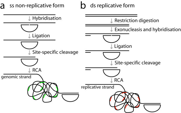
Background: In this study we utilized padlock probes and rolling circle amplification as a mean to detect and study the replication of porcine circovirus type 2 (PCV2) in cultured cells and in infected tissue. Porcine circovirus type 2 is a single-stranded circular DNA virus associated with several severe diseases, porcine circovirus diseases (PCVD) in pigs, such as postweaning multisystemic wasting syndrome. The exact reason and mechanisms behind the trigger of PCV2 replication that is associated with these diseases is not well-known. The virus replicates with rolling circle replication and thus also exists as a double-stranded replicative form.

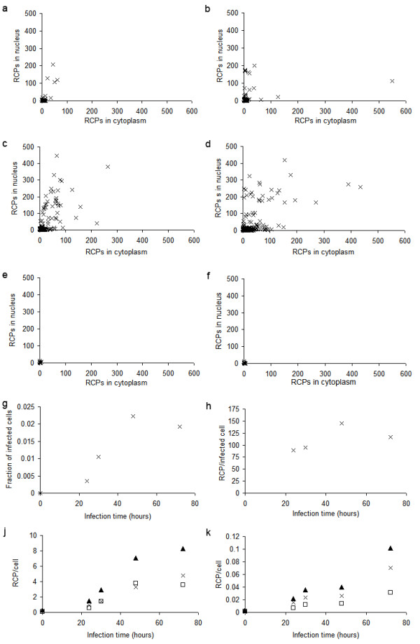
Results: By applying padlock probes and rolling circle amplification we could not only visualise the viral genome but also discriminate between the genomic and the replicative strand in situ. The genomic strand existed in higher numbers than the replicative strand. The virus accumulated in certain nuclei but also spread into the cytoplasm of cells in the surrounding tissue. In cultured cells the average number of signals increased with time after infection.

Conclusions: We have developed a method for detection of both strands of PCV2 in situ that can be useful for studies of replication and in situ detection of PCV2 as well as of DNA viruses in general.


Figures

Figure 1
Figure 1
Schematics over the padlock probe and rolling circle amplification procedure. Strand-specific padlock probes are hybridised and ligated a) directly on single stranded DNA (for the genomic form), and b) after restriction digestion and exonucleolysis on double stranded DNA (for the replicative form). The target DNA is then cut site-specifically using the combined action of MutY and EndoIV enzymes to create a free 3'-end as a starting point for RCA. After RCA the rolling circle products are visualised by hybridisation of fluorescently labelled detection oligonucleotides, green for the products from the probe specific for the genomic strand and red for the probes specific for the replicative form.
Figure 2
Figure 2
PCV2 Infection in PK-15A cells. Path of infection in PCV2 infected PK-15A cells after a) 24 hours, b) 30 hours, c) 48 hours, d) 72 hours, e) 72 hours unligated padlock probe and f) uninfected for 24 hours. Rolling circle products from the genomic strand are seen as green dots, signals from the replicative strand as red dots, and nuclei are counterstained with DAPI (blue).
Figure 3
Figure 3
Quantification of PCV2 infection in cells. PCV2 infection in PK-15A cells. a-f) Distribution of virus strand RCPs in nucleus and cytoplasm in individual cells at a) 24 h, b) 30 h, c) 48 h, d) 72 h, and e) 0 h after infection. f) negative control without ligase at 72 h after infection, g) fraction of cells with at least 15 RCPs in the nucleus, h) average number of RCPs in cells with more than 15 RCPs in the nucleus, j) average number of RCPs in nucleus are seen as white squares, in cytoplasm as crosses and totally per cell as black triangles from the padlock probe targeting the genomic strand and, k) average number of RCPs in nucleus are seen as white squares, in cytoplasm as crosses and totally per cell as black triangles from the padlock probe targeting the replicative strand.
Figure 4
Figure 4
PCV2 detected in cells with mitochondrial staining. PCV2 detected in cells with mitochondrial staining. a) Merged image with RCP detecting virus as green dots, replicative strand as blue dots and mitochondria labelled with Mitotracker seen in red, b) Mitotracker, c) signals for the genomic strand and d) signals for the replicative strand.
Figure 5
Figure 5
Detection of PCV2 in lymph node tissue. Detection of PCV2 with padlock probes in lymph node tissue from a) an experimentally infected pig, b) a natural case of PMWS and c) PCV2 negative tissue. Rolling circle products detecting the genomic strand is seen as green dots, the replicative strand is seen as red dots and the nuclei are counterstained with DAPI (blue).

References

    1. Nilsson M. et al.Padlock probes: circularizing oligonucleotides for localized DNA detection. Science. 1994;265:2085–2088. doi: 10.1126/science.7522346. - DOI - PubMed
    1. Fire A, Xu SQ. Rolling replication of short DNA circles. Proc. Natl. Acad. Sci. USA. 1995;92:4641–4645. doi: 10.1073/pnas.92.10.4641. - DOI - PMC - PubMed
    1. Lizardi PM, Huang X. et al.Mutation detection and single-molecule counting using isothermal rolling-circle amplification. Nat Genet. 1998;19(3):225–232. doi: 10.1038/898. - DOI - PubMed
    1. Allalou A, Wahlby C. BlobFinder, a tool for fluorescence microscopy image cytometry. Comput Methods Programs Biomed. 2009;94(1):58–65. doi: 10.1016/j.cmpb.2008.08.006. - DOI - PubMed
    1. Larsson C, Koch J. et al.In situ genotyping individual DNA molecules by target-primed rolling-circle amplification of padlock probes. Nat Methods. 2004;1(3):227–232. doi: 10.1038/nmeth723. - DOI - PubMed

Publication types

LinkOut - more resources